Case Report

Familial achalasia with an autosomal dominant pattern of inherence: Report of a Vietnamese family

Duc Trong Quacha,*, Yuji Urabeb, Toru Hiyamac
Author Information & Copyright
aDepartment of Internal Medicine, University of Medicine and Pharmacy at Hochiminh City, Ho Chi Minh, Vietnam
bDivision of Regeneration and Medicine Center for Translational and Clinical Research, Hiroshima University Hospital
cHealth Service Center, Hiroshima University, Higashihiroshima, Japan
*Address correspondence to Assoc. Professor Duc Trong Quach, MD., PhD. at the Department of Internal Medicine, University of Medicine and Pharmacy at Hochiminh City, 217 Hong Bang Street, District 5, Ho Chi Minh, Vietnam; Tel: +84-838-554-269; Fax: +84-839-506-126; E-mails: drquachtd@ump.edu.vn.

© Copyright 2019 MedPharmRes. This is an Open-Access article distributed under the terms of the Creative Commons Attribution Non-Commercial License (http://creativecommons.org/licenses/by-nc/4.0/) which permits unrestricted non-commercial use, distribution, and reproduction in any medium, provided the original work is properly cited.

Received: Jul 01, 2019; Revised: Aug 25, 2019; Accepted: Sep 12, 2019

Published Online: Sep 30, 2019

Abstract:

Current pathophysiologic knowledge of achalasia suggests the important involvement of genetic predisposition. However, familial achalasia is very rare and most of the case reports in literature have shown an autosomal recessive pattern of inherence. We hereby report a case of familial achalasia with autosomal dominant pattern of inherence affecting ten members in three generations of a Vietnamese family.

Keywords: Achalasia; autosomal inherence; familial achalasia; Vietnamese

1. INTRODUCTION

Achalasia is a rare esophageal disorder. Although etiology of the disease is still largely unknown, its resultant abnormalities have been well understood which include loss of esophageal peristalsis and relaxation failure of the lower esophageal sphincter [1]. Current pathophysiologic knowledge of achalasia suggests the important involvement of genetic predisposition [2-4]. However, there are few studies on familial achalasia in the literature. Most of the case reports are small with the average number of patients from 2 to 4, mostly in siblings than in members of different familial generations.

To the best of our knowledge, there are only very few case reports on familial achalasia with classical autosomal inherence pattern reported in Europe and the United States [5-7]. We hereby report a Vietnamese family with 10 affected members in three generations. This is the first such a case of familial achalasia with classical autosomal inherence pattern reported in Vietnam.

2. CASE REPORT

Family member (proband)

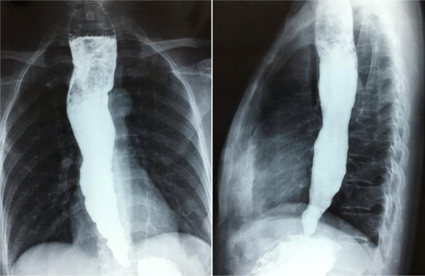
A 50-year-old female patient was referred to the University Medical Center, Hochiminh City, Vietnam because of worsening dysphagia. She had dysphagia for 20 years, initially for solid foods but subsequently for liquids. Upper gastrointestinal endoscopy showed significantly dilated esophagus with retained secretions. A high pressure zone was felt and a “given” feeling was experienced as the scope passed the gastroesophageal junction. There was no true stricture or neoplasm. Achalasia was suspected and upper gastrointestinal series were performed which confirmed the diagnosis (Figure 1). Endoscopic pneumatic balloon dilatation was performed successfully.
mpr-3-2-25-g1
Figure 1. Conventional barium esophagram. Dilated proximal esophagus as a result of the poor relaxation and opening of the sphincter (patient III1)
Download Original Figure
Family description

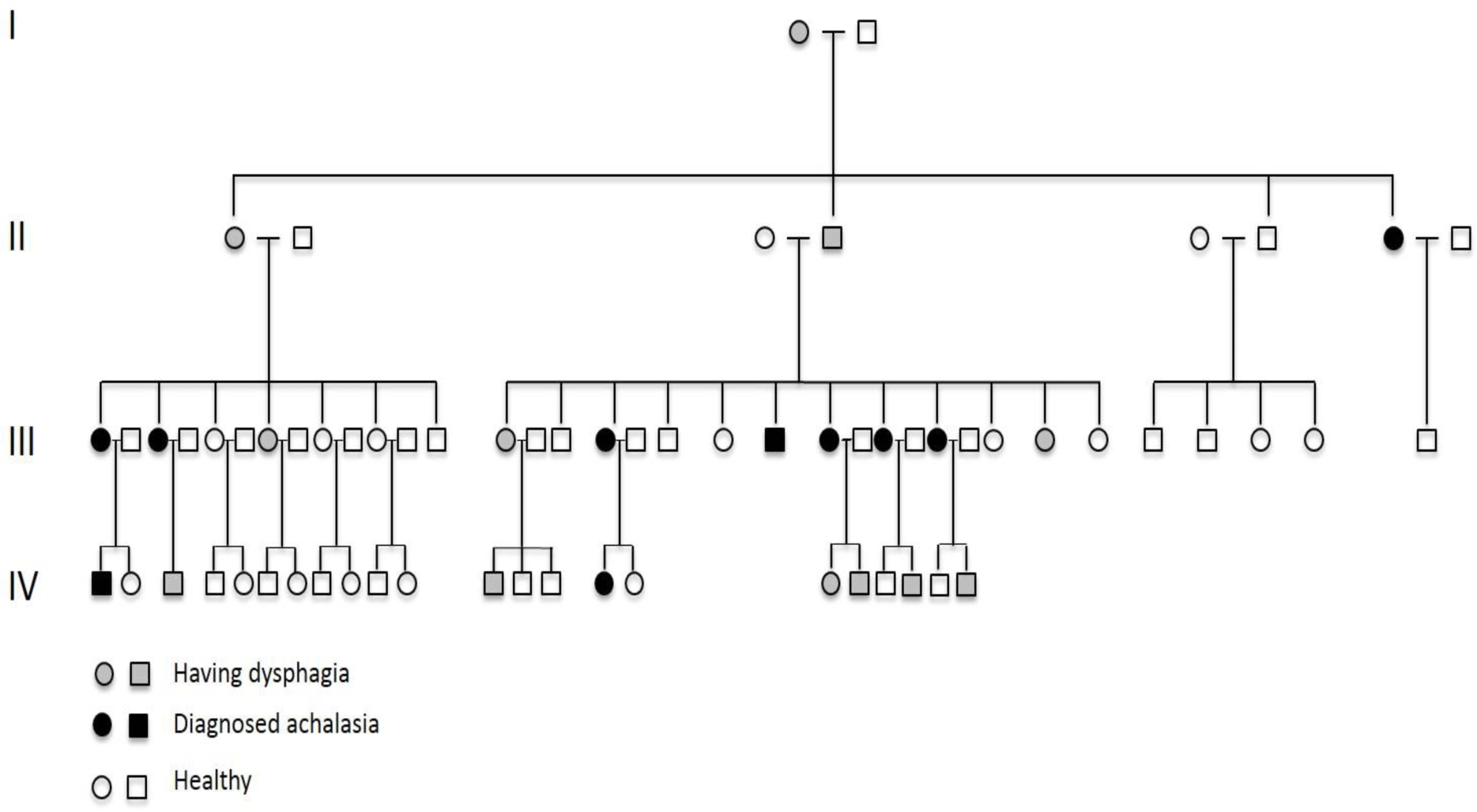
This is a four-generation family. There are ten patients (two male, 8 female) belonging to three different generations with confirmed achalasia (Figure 2). The diagnosis was made based on dysphagia, typical esophagram of achalasia and upper gastrointestinal endoscopy (to exclude other organic causes of dysphagia) at two tertiary hospitals in Hochiminh City, Vietnam (Nguyen-Tri-Phuong hospital and University Medical Center). Esophageal manometry was not performed as the equipment was not available in Vietnam. Because of different management strategies at the two hospitals, six patients admitted to Nguyen-Tri-Phuong hospital were performed Heller’s myotomy surgery while the other four patients admitted to University Medical Center were performed endoscopic pneumatic balloon dilatation. One patient with initially unsuccessful balloon dilatation was managed by myotomy surgery. In addition, there are 12 members of the family belonging to 4 different generations suffering from dysphagia. All of the family members who have been diagnosed with achalasia or suffered from dysphagia but not yet been confirmed with achalasia reported that they firstly had dysphagia at the age of 15 to 25 (Table 1). There has been no esophageal carcinoma diagnosed in this family.
mpr-3-2-25-g2
Figure 2. Pedigree of the family showing affected members and vertical pattern of inherence
Download Original Figure
Table 1. Clinical data of the ten family members with a confirmed diagnosis of achalasia
Family member Sex Beginning of symptoms (years) Age at diagnosis (years) Diagnostic method Treatment method
II7 Female 15 – 20* 59 Symptomatology BE, UGIE Pneumatic dilatation
III1 Female 15 – 20* 50 Symptomatology BE, UGIE Pneumatic dilatation
III3 Female 15 – 20* 46 Symptomatology BE, UGIE Pneumatic dilatation
III17 Female 15 – 20* 54 Symptomatology BE, UGIE Myotomy surgery
III21 Female 15 – 20* 46 Symptomatology BE, UGIE Myotomy surgery
III22 Female 15 – 20* 45 Symptomatology BE, UGIE Myotomy surgery
III24 Female 15 – 20* 42 Symptomatology BE, UGIE Myotomy surgery
III26 Female 15 – 20* 40 Symptomatology BE, UGIE Myotomy surgery
IV1 Male 25 28 Symptomatology BE, UGIE Pneumatic dilatation
IV15 Female 20 30 Symptomatology BE, UGIE Myotomy surgery

* Dysphagia onset was roughly estimated by patients as they were unable to remember the exact age of onset. BE: Barium esophagram; UGIE: upper gastrointestinal endoscopy, F: female, M: male.

Download Excel Table

3. DISCUSSION

To diagnose achalasia in the early stage of the disease, the guideline of the American College of Gastroenterology recommended to use esophageal manometry as the primary examination; and barium esophagram and upper gastrointestinal endoscopy were considered as complementary tests [8]. However, this guideline also addressed that the role of manometry is supportive to confirm the diagnosis in patients with classic endoscopic and esophagram findings. Although esophageal motility testing was not available in Vietnam at the time these patients were managed, patients with achalasia diagnosis in this family were in late stages with typical esophagram findings (esophageal dilation and peristalsis, narrow gastroesophageal junction with poor emptying of barium) and endoscopic findings (esophageal dilatation, narrow gastro-esophageal junction with no evidence of cardiac tumor) and, therefore, the definite diagnosis of achalasia could be reliably established. Affected members in the fourth generation of this family were diagnosed and received treatment at younger ages compared with those in the older generations as a consequence of the awareness of this disease.

Most of the cases of familial achalasia presented in literature showed an autosomal recessive pattern of inherence. A large prospective cohort study over the last 24 years in Nigeria recently reported 18 families with familial achalasia [9]. All patients in these families were observed in siblings, and consanguineous marriages were common among parents of these patients. There were very few case reports on classical autosomal inherence pattern in the literature. One case is a Southern American family with six affected members in three generations. The beginning age with symptoms was about 22 – 25, similar to patients in our case report. Two members finally died because of esophageal carcinoma in their late seventies and eighties [6]. Another case is an Italian family with five affected members in three generations. The beginning age with symptoms was not mentioned and there was no esophageal carcinoma occurred during the follow-up period [5]. The number of members with confirmed achalasia in our case report was the largest among the three case reports. In addition, 12 members belonging to four generations suffer from mild dysphagia. The subjects have not consulted or already consulted but have not been confirmed with achalasia. If motility testing were available, there would have been more cases with achalasia diagnosed among these subjects. The pedigree of this family clearly showed a vertical transmission of the disease and strongly suggested the autosomal dominant pattern of inherence (Figure 1).

In genetically predisposed subjects, it is suggested that environmental factors may trigger an autoimmune antibody response against myenteric plexus of the esophagus [10]. In fact, a recent large case-control study reported that patients are more often affected by viral infections before achalasia onset, most significantly for varicella-zoster virus infections [11]. In addition, this study also showed that achalasia can also be triggered by pregnancies in female HLA-DQß1 insertion carriers. These findings help to explain the dysphagia onset around the age of 15 – 25 years and the female predominance among affected members in our presented family. Recent studies reported that allergic and autoimmune disorders are comorbid disease conditions in patients with achalasia [12, 13]. However, we have not identified any significant comorbid conditions in members of this family. In addition, no cases with esophageal cancer have been diagnosed in this family until now. However, patients diagnosed with achalasia in our case report are still at much younger ages compared to those with esophageal carcinoma in the first case report [6]. Therefore, the development of this malignancy needs to be further followed-up.

In conclusion, we report for the first time familial achalasia with autosomal dominant pattern of inherence in Vietnam and review the literature. Future genetic study is required to identify the causative gene.

ACKNOWLEDGMENTS, FUNDING, AND DISCLOSURES

This case report with the genetic analysis has been presented during the 5th NGS site meeting, which was a closed genetic meeting in Japan. In this meeting, Dr. Urabe consulted the NGS result to other researchers. The number of identified mutations was 19343 mutations, which was narrowed down to 6 candidate mutations. As a result of the consultation, it is impossible to announce the genetic data publicly at the present and long-term follow up is required to have more information to identify the causative mutation. Therefore, we have excluded researchers who were associated with the genetic analysis from the co-authors’ list and this case report in this current version is only with clinical presentation.

Notes

Author contributions: Duc Trong Quach collected data. Duc Trong Quach and Yuji Urabe prepared the manuscript. Toru Hiyama supervised and revised the preparation of the manuscript. All authors have read and accepted the final version of this manuscript.

Notes

Financial support: None.

Notes

Conflicts of interest: None.

Notes

Ethics approval and consent to participate: Not applicable.

Notes

Consent for publication: Written informed consent was obtained from the patient for publication of this case report and any accompanying images. A copy of the written consent is available for review by the Editor-in-Chief of this journal.

REFERENCES

1.

Reynolds JC, Parkman HP. Achalasia. Gastroenterology clinics of North America. 1989 Jun; 18((2)):223-55

2.

Gockel I, Becker J, Wouters MM, Niebisch S, Gockel HR, Hess T, et al. Common variants in the HLA-DQ region confer susceptibility to idiopathic achalasia. Nature genetics. 2014 Aug; 46((8)):901-4

3.

Singh R, Ghoshal UC, Misra A, Mittal B. Achalasia Is Associated With eNOS4a4a, iNOS22GA, and nNOS29TT Genotypes: A Case-control Study. Journal of neurogastroenterology and motility. 2015 Jul; 3021((3)):380-9

4.

Wouters MM, Lambrechts D, Becker J, Cleynen I, Tack J, Vigo AG, et al. Genetic variation in the lymphotoxin-alpha (LTA)/tumour necrosis factor-alpha (TNFalpha) locus as a risk factor for idiopathic achalasia. Gut. 2014 Sep; 63((9)):1401-9

5.

Vailati C, Mazzoleni G, Testoni PA, Passaretti S. An Italian family with inherited achalasia. Digestive and liver disease : official journal of the Italian Society of Gastroenterology and the Italian Association for the Study of the Liver. 2013 Jun; 45((6)):524-5

6.

Gordillo-Gonzalez G, Guatibonza YP, Zarante I, Roa P, Jacome LA, Hani A. Achalasia familiar: report of a family with an autosomal dominant pattern of inherence. Diseases of the esophagus : official journal of the International Society for Diseases of the Esophagus. 2011 Jan; 24((1)):E1-4

7.

Halpern AL, Torphy RJ, McCarter MD, Sciotto CG, Glode LM, Robinson WA. A familial germline mutation in KIT associated with achalasia, mastocytosis and gastrointestinal stromal tumors shows response to kinase inhibitors. Cancer Genetics. 2019; 233-234:1-6

8.

Vaezi MF, Pandolfino JE, Vela MF. ACG clinical guideline: diagnosis and management of achalasia. The American journal of gastroenterology. 2013 Aug; 108((8)):1238-49 quiz 50

9.

Tebaibia A, Boudjella MA, Boutarene D, Benmediouni F, Brahimi H, Oumnia N. Incidence, clinical features and para-clinical findings of achalasia in Algeria: Experience of 25 years. World journal of gastroenterology. 2016 Oct; 1422((38)):8615-23

10.

Gyawali CP. Achalasia: new perspectives on an old disease. Neurogastroenterology and motility : the official journal of the European Gastrointestinal Motility Society. 2016 Jan; 28((1)):4-11

11.

Becker J, Niebisch S, Ricchiuto A, Schaich EJ, Lehmann G, Waltgenbach T, et al. Comprehensive epidemiological and genotype-phenotype analyses in a large European sample with idiopathic achalasia. European journal of gastroenterology & hepatology. 2016 Jun; 28((6)):689-95

12.

Becker J, Haas SL, Mokrowiecka A, Wasielica-Berger J, Ateeb Z, Bister J, et al. The HLA-DQbeta1 insertion is a strong achalasia risk factor and displays a geospatial north-south gradient among Europeans. European journal of human genetics : EJHG. 2016 Aug; 24((8)):1228-31

13.

Romero-Hernandez F, Furuzawa-Carballeda J, Hernandez-Molina G, Alejandro-Medrano E, Nunez-Alvarez CA, Hernandez-Ramirez DF, et al. Autoimmune comorbidity in achalasia patients. Journal of gastroenterology and hepatology. 2017 May; 31