1. INTRODUCTION
Intraorbital foreign body is an object that lies within the orbit but outside the ocular globe, usually from injuries that related to high-speed flying objects such as a bullet or industrial accidents, traffic accidents, daily life accidents… Basing on the composition, intraorbital foreign body can be classified into (a) metallic and (b) non-metallic. Non-metallic objects, in their turn, are classified into well-tolerated objects (glass, silicon) and poor-tolerated objects (vegetable matters, wood, chemicals…). Metallic and well-tolerated objects can be left alone in the orbit without causing any harm; however, poor-tolerated objects may cause persistent inflammatory reaction or forming local abcess refractory to medical treatment and need to be removed [1].
Untreated intraorbital foreign body may lead to various complications, such as orbital cellulitis, endopthalmitis, orbital abcess, or even septicaemia, threatening patient’s life [2, 3]. Not only related to structures in the orbit, intraorbital foreign body is also associated with adjacent sinuses, central nervous system, as well as large vessels of head and neck. Surgery plan is based on whether the object is magnetic or non-magnetic, location of the object in the orbit and adjacent tissues injury caused by the object, such as optic nerve compression, infection or intraocular muscles injury. In this article, we report a case large, non-metallic foreign body which localized in dangerous position and caused local abcess [4].
2. CASE REPORT
A 38-year-old male patient was admitted to Ho Chi Minh City Eye Hospital on February 13th 2018 due to blurry vision and pain on left eye (OS) after being hit by a windscreen wiper 13 hours ago. One hour after the injury, he has his eyelid sutured in a tertiary care hospital without any further imaging diagnosis and treatment. He came to our hospital 12 hours after the eyelid surgery because the pain and swelling became worse. He has no history of diabetic mellitus, hypertension, eye diseases, neurological diseases and head trauma before.
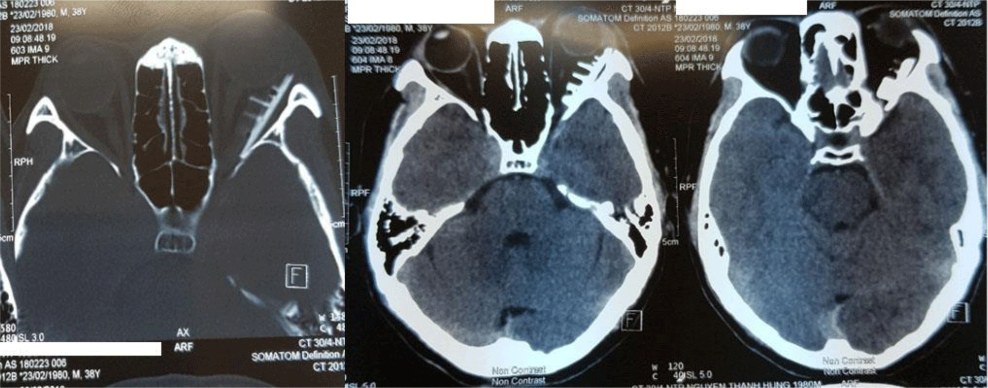
At the time of admission, the patient had 40°C fever, his best-corrected visual acuity (BCVA) on left eye was 20/60. Clinical examination on left eye revealed ptosis with sutured lid injury being red, swollen, conjunctival injection, corneal abrasion and restricted eye movements in all directions. CT-scan imaging was performed and revealed a polymorphous foreign body near the lateral wall of the orbit and adjacent to the apex, 7×10×35mm in size; grade III proptosis, swollen lateral rectus muscle and the presence of air in the orbit (Figure 1). Based on the patient history, clinical examination and CT-scan images, he was diagnosed with intraorbital foreign body and orbital cellulitis of OS. After a thorough discussion with colleagues from neurosurgery department, the patient was indicated a surgery to remove the foreign body.

After 3 days using intravenous ceftriaxone 1g q.i.d and metronidazole 0.5g q.i.d, the patient’s fever and swelling resolved. He underwent surgery under general anesthesia condition, after being explained the risk of optic nerve injury, septicaemia and meningitis. As the foreign body is quite large with many edges and near the apex, removing it may probably result in injury of the optic nerve. In order to avoid this complication, we performed lateral orbitotomy.
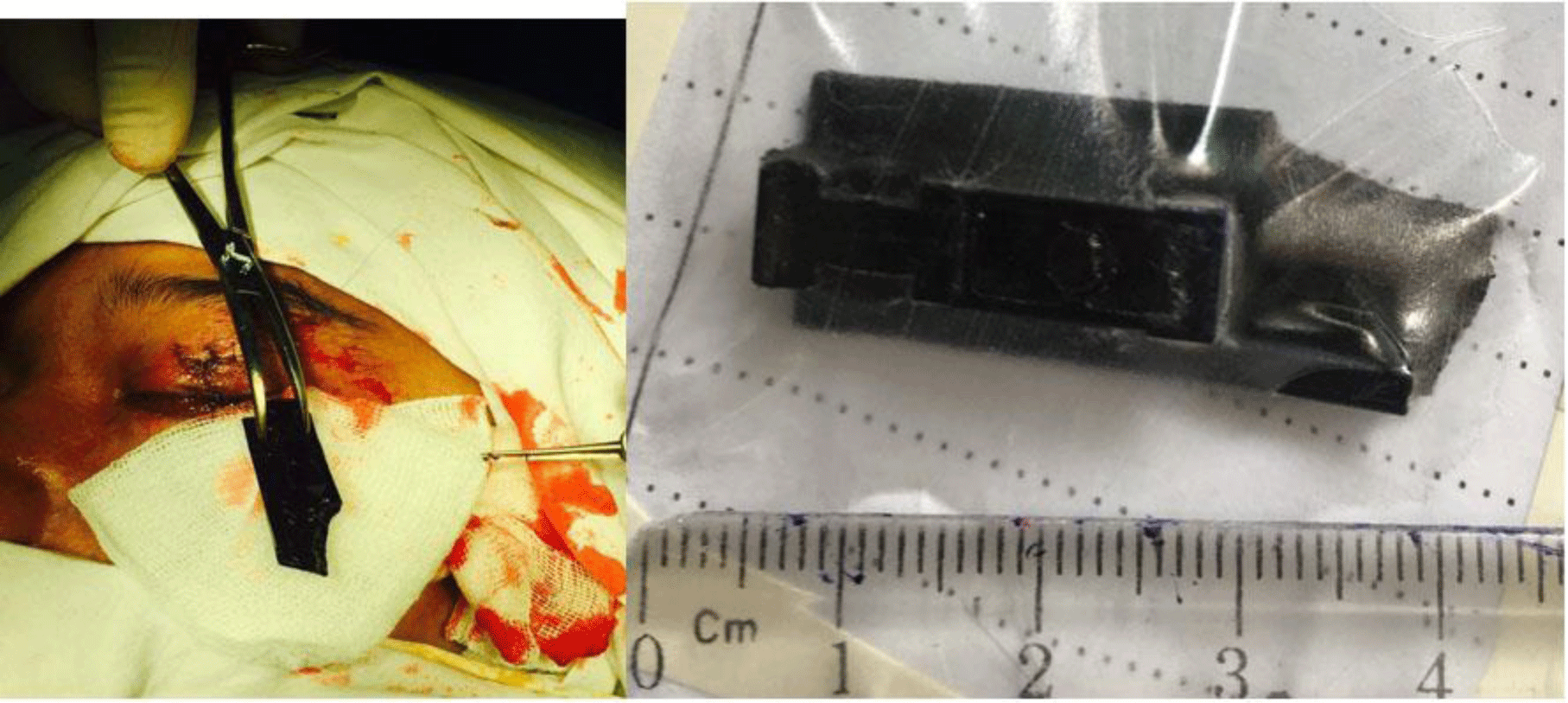
A horizontal 20mm incision was made right at the unification of the upper and lower eyelid towards the temple, soft tissues and bone membrane was dissected to expose the bone. A piece of lateral wall of the orbit, 20×30mm in size, was removed by an electric drill. Right after removing the lateral wall, we managed to approach the foreign body, gently removed it without injuring adjacent tissues. The foreign body was made of plastic, 7×10×35mm in size with many edges and adjacent to the orbital apex (Figure 2). The dissected piece of bone was then reattached, bone membrane and other soft tissues were sutured. After placing spongel and penrose, serum antitetanus was injected to the patient. After the surgery, the patient was treated with intravenous metronidazole 0.5g t.i.d, intravenous ceftriaxone 1g t.i.d, oral methylprednisone 0.5mg/kg and topical levofloxacin q.3.h. The penrose was removed 1 day after the surgery.
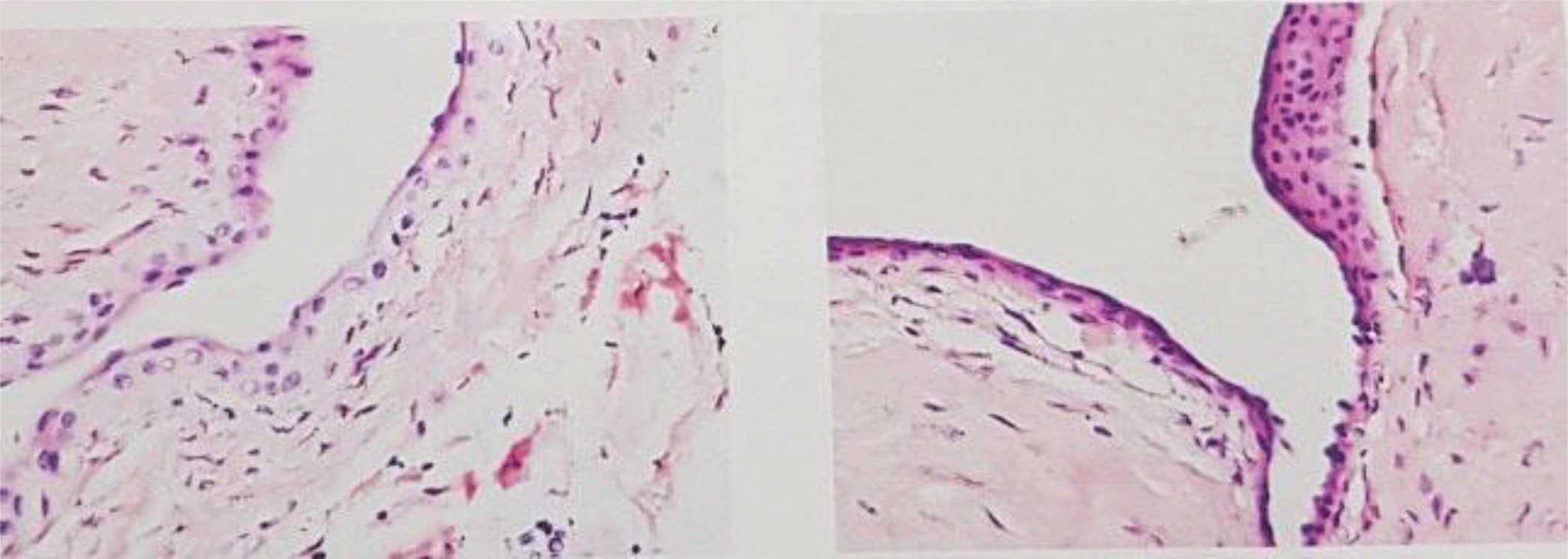
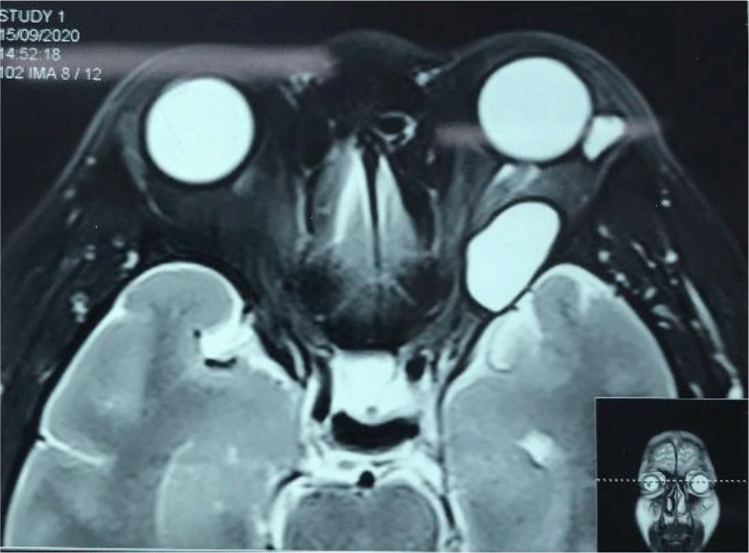
At 7 days post-op, the patient’s BCVA of OS was 20/200. At 2 weeks post-op, BCVA was 20/40 with proptosis resolved, some progression in eye movements and mild diplopia on lateral gaze. At 3 months post-op, patient BCVA was 20/30, diplopia resolved and eye movements returned to normal condition; the patient’s follow-up result remained the same until 18-month follow-up. However, at 24-month follow-up, his left eye developed mild proptosis without the presence of eye movements disorder or blurry vision. The MRI images showed both intraconal and extraconal homogenous masses with distinct bag and increased inhencement on T2, size 15×18×24mm with the intraconal mass and 7×8×10mm with the extraconal mass (Figure 3). To clarify the nature of the tumors and prevent them from enlarging, we decided to perform operation. He underwent lateral orbitotomy again to remove the tumors. The tumors were removed successfully and the pathology resulted in epithelial cysts (Figure 4). Seven days post-operation, patient BCVA was 20/20 in the OS, with normal eye movements and eyelids position.

3. DISCUSSION
Diagnosis of intraorbital foreign body faces many difficulties as (a) Patients did not usually supply a honest history right from the start, (b) Non-metallic and small foreign body may be misdiagnosed due to the absence of its image on B-scan ultrasonography and X-ray images and (c) The ophthalmologists may have a tendency to pay attention only to injuries on the ocular globe, or the foreign body may penetrate into the orbit from the globe or the adjacent sinuses, instead of an open injury on the skin [5].
In this patient, the confused trauma history (he hid some details of trauma history and only told us in detail after that) has caused difficulty in making a precise diagnosis and primary treatment. However, orbital foreign body should be suspected on a patient with lid injury, intact ocular globe and conjunctival edema. Orbital cellulitis, orbital fracture, orbital apex syndrome, trauma optic neuropathy and orbital foreign body should be excluded on a patient with pain, lid edema, conjunctival edema, restricted eye movements, proptosis, ptosis and blurry vision. A combination of careful case history, clinical manifestations and imaging tests is critical to exclude orbital foreign body [6]. Patient with ocular trauma should take B-scan ultrasound and orbital X-ray images on primary approach. An injury with the presence of orbital foreign body should be treated by specialized centers. Head CT-scan is considered gold standard in diagnosing metallic orbital foreign body. The composite of foreign body can be estimated based on the difference in contrast signal on CT-scan images. The sensitivity of CT-scan on detecting orbital foreign body is about 65% with foreign body <0.06 mm3 in size to 100% with foreign body >0.06 mm3 in size. Besides, CT-scan can detect orbital fracture and orbital infection, if present [3, 7].
Indications of surgery to remove orbital foreign body include (a) poor-tolerance foreign bodies should be removed as soon as possible to prevent infection or abcess (b) foreign bodies with restricted eye movements, optic nerve compression and decreased visual acuity. Small metal, glass or plastic foreign bodies can be left alone if no sign or symptom present [8]. With the lateral wall being 43.78mm in length, in this patient, the foreign body (35mm in length) took around 80% length the wall. With such size, removal of the foreign body is mandatory in this patient [9].
Choosing a surgical technique to approach to the foreign body is essential. In this patient, the shape and location of the foreign body made it easy to damage adjacent tissues, including the orbital apex. A large approach was necessary to prevent damage to the optic nerve and adjacent tissues; as well as the use of electric magnet was impossible because the foreign body itself was non-metallic. Therefore, lateral orbitotomy was and appropriate choice because this technique help approach deep lesions (tumors or foreign bodies) in or below bone membrane, inside or outside the muscle cone and lateral to the optic nerve.
When patient presented with fever and a foreign body near the orbital apex, neurosurgery colleagues were consulted to evaluate the damage to the brain and spead of infection to the spinal cord. As a result, we had high agreement that this was a case of orbital cellulitis with the probability of infection spreading to the brain being considerably high, especially with anaerobic bacteria. Symptoms improved by the use of intravenous ceftriaxone and metronidazole. After 1 week the fever resolved but restricted eye movements still present; after 2 weeks BCVA 20/40, mild restricted eye movements still present. After 3 months, BCVA recovered to 20/30, no restricted eye movements present.
Beside intraorbital foreign body, damage to the central nervous system and adjacent sinuses should be confirmed to combine treatment. Head CT-scan images can help confirm the diagnosis. The trauma could affect cranial fossa via orbital. Air in the cranial fossa, damage of frontal, sphenoidal and ethmoidal sinus, intracranial fluid in the nasal cavity or conjunctival edema should be inspected to confirm or exclude the damage of central nervous system [7].
Injection of serum anti tetanus and broad-spectrum antibiotics are critical to prevent infection. In the operation, the incision can be opened more widely to give better inspection and easier to remove the foreign body, or cleaning the proximal abcess. The injuries, which are more likely to bleed or form local abcess, can be left open or dried with a penrose [8].
At 24-month follow-up, the patient developed epithelial cysts in the same side with the previous surgery. It was assumed that these serous cysts resulted from the reaction of adjacent lymphoid organs after the previous surgery. These cysts, however, did not affect either visual acuity or eye movements and were successfully removed.
This is a rare case of large and complicated intraorbital foreign body, with follow-up time long enough to know what might happen after lateral orbitotomy. However, a considerable limitation of this study is that we do not have many cases to know the prevalence of secondary serous cysts after surgery.
Conclusion
In order to diagnose early and treat properly a case of intraorbital foreign body, taking history and mechanism of trauma carefully and CT-scan images are essential. Besides, the aid of CT-scan images of the orbit, the globe, the sinuses and the cranial bones are necessary to locate precisely the foreign body and to find out injuries of adjacent structures caused by the foreign body. After discovering size, structure and location of the foreign body, appropriate treatment strategy can be sorted out. Treatment outcome depends on the early diagnosis, skill and experience of the surgeon to minimize secondary injuries and the risk of incomplete removal of foreign body. High dose antibiotics are critical to prevent infection.


