1. INTRODUCTION
In 2002, PEComas were defined as “mesenchymal tumors composed of histologically and immunohistochemically distinctive perivascular epithelioid cells” by The World Health Organization (WHO) [10]. Later in 2013, this family of neoplasm was defined by WHO as mesenchymal tumors that usually express melanocytic and smooth-muscle markers. PEComas family includes angiomyolipoma (AML), clear cell sugar tumor of lung (CCST), lymphangioleiomyomatosis (LAM)… [1]. PEComas can arise in many organs; and uterus is the most common site. Hepatic PEComas are extremely rare [3]. Moreover, it is very difficult to draw correct diagnosis of PEComas clinically because it is often misdiagnosed with other tumors [2]. We report here a case of primary hepatic PEComa occurring in 44-year-old woman with literature reviews.
2. CASE REPORT
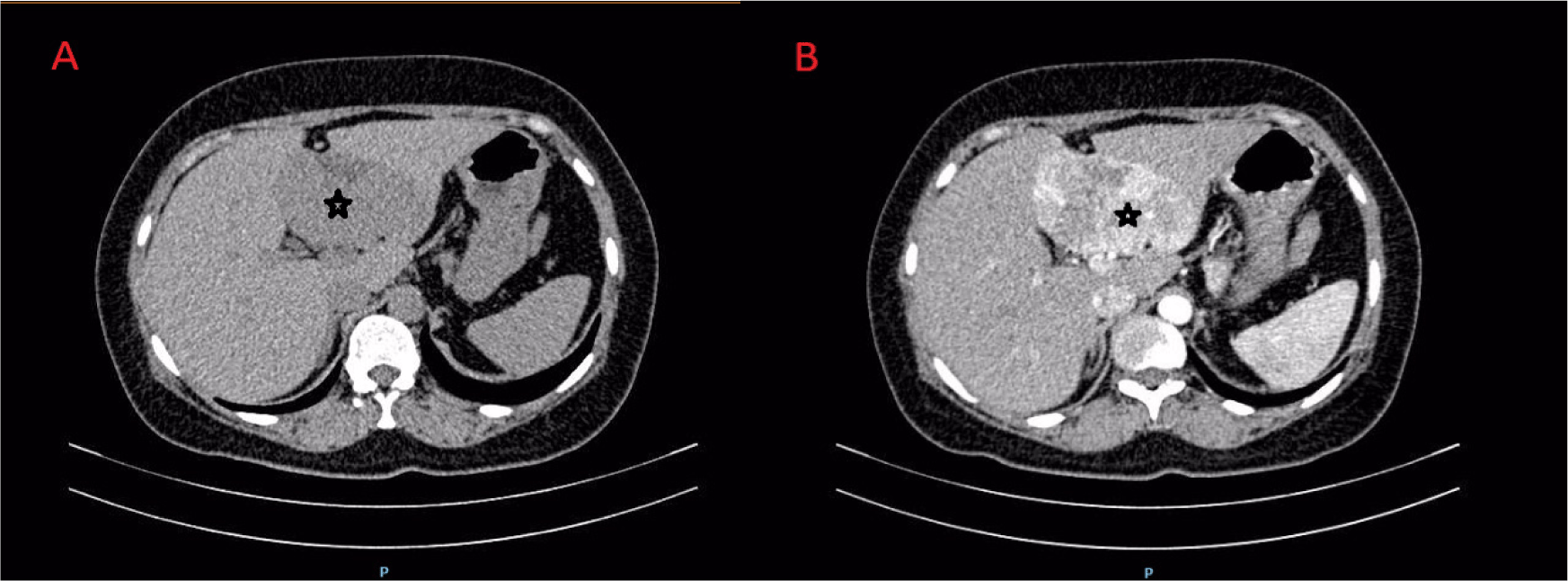
In February 3th, 2017, a 44 years old woman came to University Medical Center for Health check up and performed abdominal ultrasonography. She was accidentially detected a liver tumor. Laboratory test revealed a normal liver function. CT Scan highlighted a hypervascular lesion in left liver which can not exclude hemangioma, adenoma, AML... (Figure 1).

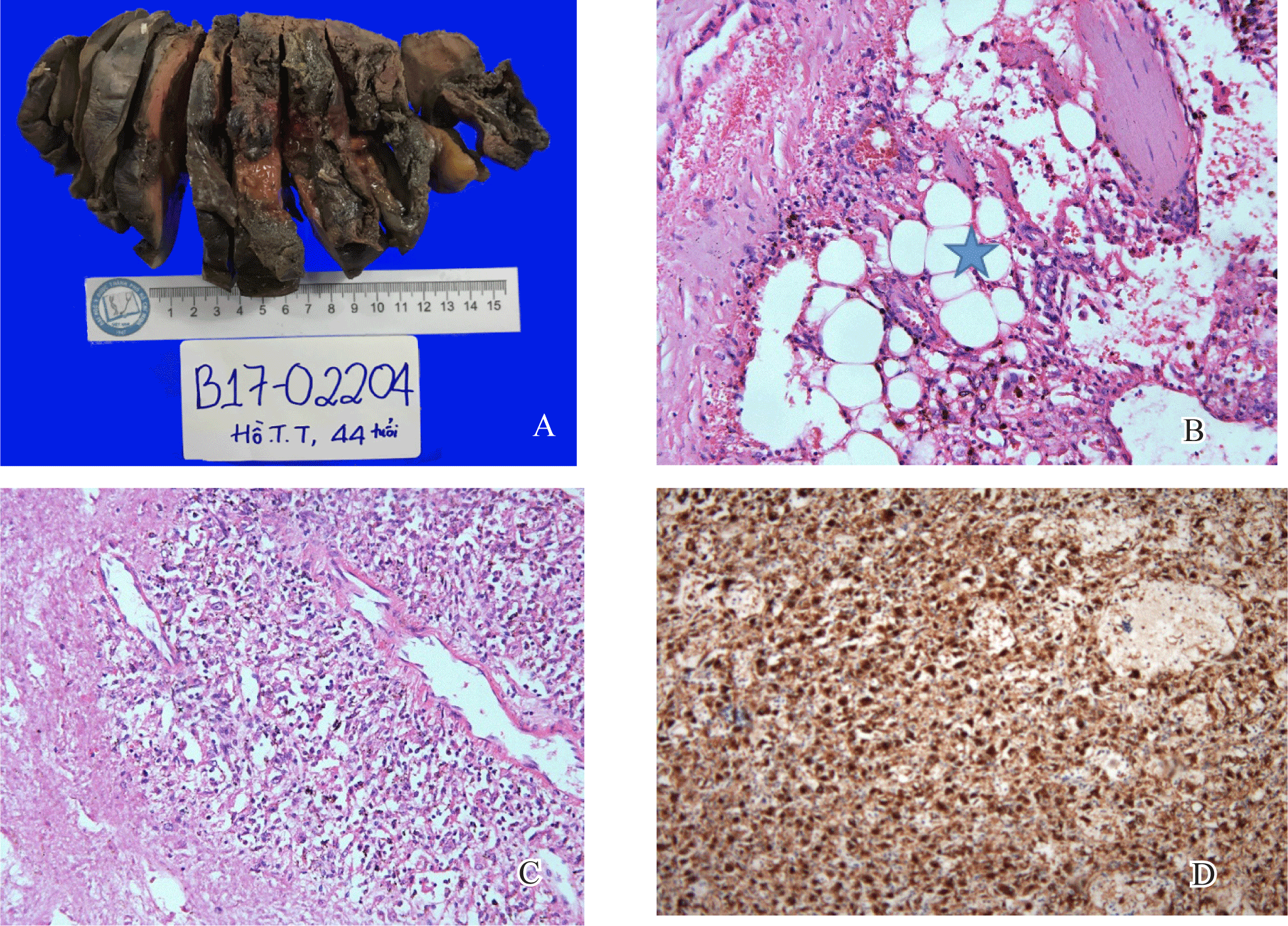
Tumor was measured 6x4 cm in dimensions, relatively well-demarcated, not encapsulated with red cut surface (Figure 2).

The tumor mainly composed of epithelioid cells, which had abundant granular eosinophilic cytoplasm, eccentrically located round nuclei with small nucleoli, and foci of mild to moderate nuclear atypism. Mitotic figure or necrosis was not observed. The nest or trabecular patterns of tumor cells were surrounded by thin-walled capillary vessels as well as thick and hyalinized blood vessels. Adipose tissue is also predominant. Scatter foci of extramedullary hematopoiesis was recognized. Foam cells and melanin-like brown pigments sparsely appeared in the sinusoidal spaces between the tumor cells. The tumor cells are strongly and diffusely immunoreactive for melanotic marker (HMB45) and negative with Desmin (Figure 2).
3. DISCUSSION
PEComa is a novel terminology for mesenchymal tumor with feature of perivascular epithelioid cell which was defined by WHO in 2002. The most common primary sites of PEComa at presentation are the uterus, vulva, rectum, heart, breast, urinary bladder... [1]. Although PEComas are commonly asymptomatic, they may present with vague pain. Hepatic PEComa is rather rare with clinical characteristics similar to other hepatic primary tumor; therefore, it is often misdiagnosed with other tumors [2]. The enhanced and drainage imaging patterns of hepatic PEComas are mimic those of hepatocellular carcinoma (HCC). Contrast enhanced ultrasonography showed early influx of the contrast agent into the tumor and rapid drainage of arterial blood to veins. In addition, power Doppler ultrasonography, which revealed displaced vessels around the tumor [2]. On CT scan, most neoplasms present as hypo intense with significant enhancement on arterial phase. The portal phase was variable. Almost all lesions reported show low-signal on T1-weighted images, high-signal on T2-weighted images [4]. Hepatic PEComa should be distinguished from focal nodular hyperplasia (FNH), hemangioma, adenoma, and other hepatic tumors [5].
In our case, hepatic tumor appeared hypervascular with low enhancement during venous phase, which is so challenging in differential diagnosis with HCC. The portal phase was variable. Almost all lesions reported show low- signal on T1-weighted images, high-signal on T2-weighted images. These radiologic findings can be confused, most often, with those of hepatocellular carcinoma or simple haemangioma [4]. In a study of Tan Y et al. ,3/7 cases were diagnosed HCC preoperatively, 2/7 cases were diagnosed FNH preoperatively and 2 cases without preoperative diagnosis [2]. Diagnosis of PEComa on CT Scan can rely on fat tissue, but of 12 cases reported by Tay S et al., 5 cases revealed a well-demarcated mass without fatty density on CT scans [5]. Liver adenoma and HCC also have fat compartment. Owing to that, morphology plays an important role in making a correct diagnosis of PEComa.
Grossly, tumor is usually found single, with median size about 8 cm in diameter, well-demarcated, without a capsule. Cut surface appear yellow or brown depending on the component of fat tissue. Necrosis and hemorrhage may be found in large tumor [1]. Tumor cells appear epithelioid, which have abundant granular eosinophilic cytoplasm, distinct cell border, eccentrically located round nuclei with small nucleoli. Tumor cells are arranged in dense sheets as epithelial-like pattern with abundant dilated vascularity [1]. These tumors are positive for HMB45, Melan-A, S-100, and SMA, but negative for CgA, Syn, CK, CD117, CD10, and CD34 [1]. In our case, the tumor cells are negative for Desmin. Folpe AL et al. showed that only 8/22 cases of PEComa positive with Desmin, while HMB-45 was the most specific marker for PEComa disease [8]. To archive final diagnosis, the pathologists need to mention the present of adipose tissue, as well as thick-walled arterial or thin-walled venous-like spaces for not confusing with other primary liver mesenchyme tumors or GIST and melanoma liver metastasis.
The histopathologic differential diagnoses of PEComa are quite numerous and influenced by the tumor sites and morphology. Our present case was consistent with monomorphic variants of PEComas, composed almost exclusively of epithelioid cells and with blood vessels and adipose tissues. For confirmation, immunohistochemical stains for HMB45 or Melan A should be performed. Moreover, in our case, foci of extramedullary hematopoiesis were also observed. In our case, the tumor cells were diffusely and strongly immunoreactive for HMB45 but not for Desmin. Metastatic clear cell carcinoma from kidney or adrenal glands could be excluded from the absence of the tumor in the primary sites.
The origin of tumor is still a dispute. As hypothesis, the tumor comes from neural crest, another tells that the origin is perivascular smooth muscle or myoblast [7]. PEComas can display characteristics of both benign and malignant tumors and the primary treatment is resection. PEComas displaying any combination of infiltrative growth, marked hypercellularity, high nuclear grade and hyperchromasia, high mitotic activity (> 1 mitotic figure I 50 HPF), atypical mitotic figures, and coagulative necrosis should be regarded as malignant [8]. Tumor size > 5 - 8 cm is also one of poor prognostic factors of PEComa [8]. According to Folpe AL et al, recurrence and/or metastasis of PEComa was strongly related to tumor size, mitotic activity, and necrosis [8].
4. CONCLUSION
PEComa is reported in many organs, but rarely in liver. Preoperative diagnosis of hepatic PEComa is challenging, so that the role of pathology is very important to draw final diagnosis. PEComa has exclusive morphology and coexpression with muscle and melanocytic markers. PEComa is not always benign; Small PEComas without any worrisome morphological features are most likely benign.


