1. INTRODUCTION
Since being recognized as a global pandemic, the novel Coronavirus Disease 2019 (COVID-19) is still a leading medical concern both globally and in Vietnam [1] [2], especially when more and more SARS-CoV-2 variants appear with substantially faster spread [3]. In field hospitals, identifying high-risk patients based on clinical features is a top priority to prevent catastrophic development to COVID-19 patients. Early identification of individual phenotypes is essential in patients who are particularly inclined to develop severe COVID-19 and who may need hospital admission [4, 5]. In order to identify individuals or subgroups of patients with COVID-19 pneumonia who are at higher risk, and to be better prepared for disease progression and consequences, detailed characterization of patients with COVID-19 pneumonia is required. More recently, several meta-analyses have found that obesity, as measured by BMI and non-BMI indicators, may be a significant contributing factor to poor COVID-19 prognosis, resulting in a decline in survival outcomes among COVID-19 hospitalized patients [6] [7] [8].
Chest imaging proved crucial in the early diagnosis and treatment of persons with suspected or confirmed respiratory infections of COVID-19 during the SARS-CoV-2 pandemic. Although chest computed tomography (CT) is considered the gold standard approach for detecting pulmonary abnormalities, it is an inaccessible and nonreproducible tool, particularly in makeshift field hospitals in developing countries. As a consequence, chest X-ray (CXR) has emerged as the primary imaging modality for early clinical treatment and severity evaluation [9] [10]. Several CXR scoring systems have been developed to stratify the severity of lung injury in CXR in COVID-19 patients and have been introduced to different clinical settings, allowing early risk assessment of COVID-19 patients at the time of admission [9] [10] [11]. Furthermore, a higher CXR severity score has been associated with in-hospital mortality [12]. Among these scoring systems, the Brixia score, which was proposed for COVID-19 patients specifically, is one of the most widely used scoring systems in many countries [13] [14] [15]. Furthermore, many studies have shown that obese individuals had a higher risk of severe grade CXR due to COVID-19 pneumonia than those of normal weight [16]. In patients with COVID-19, a high CXR severity score and a high BMI can be significant risk factors for hospitalization and intubation. [9]. In patients with COVID-19, a linear connection has been documented between BMI and the degree of lung damage on chest radiographs [7] [8] [16]. However, in Vietnam, the BMI was categorized with Asian criteria was limited. Furthermore, owing to resource restrictions and a lack of radiologist expertise at the field hospital, non-radiologist practitioners are responsible for physical examination and CXR interpretation to make clinical decisions. As a result, additional study is needed to investigate the relationship between BMI and the radiographic severity of lung damage in Vietnamese patients who attended primary care facilities. The goal of this study was to investigate if there was a correlation between BMI-based obesity (as defined by the Asian-BMI classification) and the severity of lung damage (as measured by the Brixia score) in hospitalized COVID-19 patients.
2. MATERIALS AND METHOD
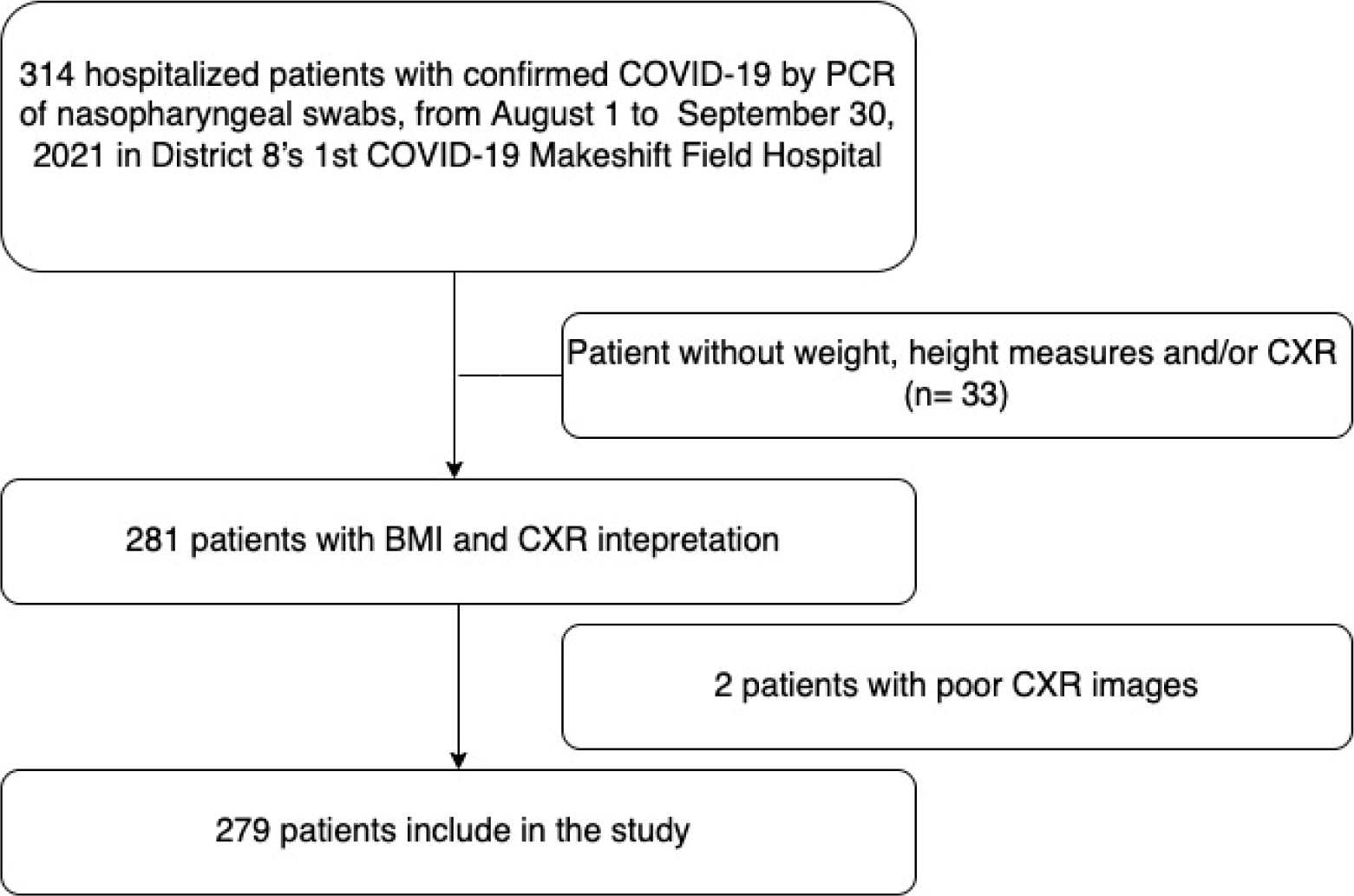
The retrospective study is based on medical records of COVID-19 patients from District 8’s 1st COVID-19 Makeshift Field Hospital, Ho Chi Minh City, Vietnam, from September 1, 2021, to September 30, 2021. Inclusion criteria were the following: (a) a final diagnosis of COVID-19 infection confirmed by a real-time reverse transcriptase-polymerase chain reaction (RT-PCR) of nasopharyngeal swabs and (b) an initial CXR performed within 24 hours after hospitalization. Exclusion criteria included patients under the age of 18 years, a history of pulmonary tuberculosis, pneumonia, and lung cancer. The flowchart of study was shown in Figure 1.

When patients with COVID-19 infection were admitted to our hospital, basic medical information such as demographic, anthropometric data, history of medical conditions, vital signs, and laboratory tests was added to the standardized medical documents. The necessary data for this study were gathered. The extracted data was composed of age, gender, BMI, history of medical conditions, and vital signs, including blood pressure, heart rate, temperature, and saturation of pulse oxygenation (SpO2). BMI was documented upon admission, either by self-report of obtainment by nursing staff and was classified according to Asian criteria: underweight (BMI < 18.5), normal (18.5 ≤ BMI ≤ 22.9), overweight (23.0 ≤ BMI ≤ 24.9), and obese (BMI > 25) [17].
The Ethics Council approved the study in Biomedical Research at the University of Medicine and Pharmacy at Ho Chi Minh City on November 12, 2021 (approval number 603/DHYD-HDDD). Informed consent was surrendered due to the retrospective disposition of this study. The patient’s identification was shielded by coding them with anonymous designated codes.
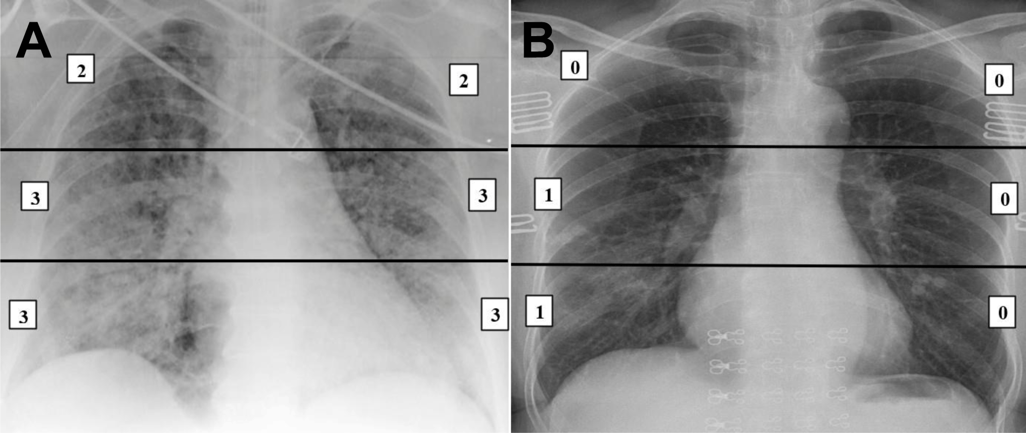
The severity of lung injury on CXR was classified by Brixia score [11]. Two internal medicine physicians independently reviewed the Brixia chest scoring system and later agreed on a severity difference. Brixia score is obtained by dividing the lungs into six regions on a CXR: (i) superior (I and IV): above the lower border of the aortic arch, (ii) midway (II and V): below the inferior border of the aortic arch and above the lower border of the right inferior pulmonary vein (i.e., hilar-hilar structure), and (iii) inferior area (III and VI): below the inferior border of the lower right pulmonary vein (i.e., lung bases). Scores (from 0 to 3) were calculated for each region based on types of pulmonary infiltration, as follows: Score 0 - no lung involvement; Score 1 - interstitial infiltration; Score 2 - interstitial and alveolar infiltration (interstitial predominance); Score 3 - interstitial and alveolar infiltration (alveolar predominance). The scores of the six lung regions were added together to obtain a total radiographic severity score ranging from 0 to 18 points. The scoring system did not include other chest radiographic features (e.g., pleural effusion, pulmonary vascular enlargement). Figure 2A and B reveals how to score each region in the Brixia score of two patients in our study.

Table 1 would present the form of the mean (standard deviation) if the continuous variables were normal and median (min, max) for non-normality continuous variables. Moreover, categorical variable summaries in the form of a number (%).
The number of Brixia scoring systems per CXR film was modeled using Poisson regression with predictors of gender and BMI. The probability of Brixia score given the gender and BMI:
Note:
βi : regression coefficient
All statistical analyzes were performed using R Statistical Software (version 4.1.0; R Foundation for Statistical Computing, Vienna, Austria). The p-value < 0.05 was considered statistically significant.
3. RESULTS
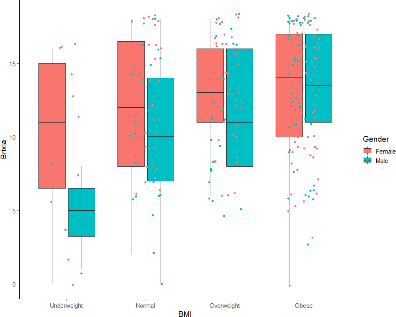
Our study was carried out with a total of 279 patients with COVID-19 (118 men and 161 women). Table 1 describes the baseline characteristics of the patients. The median age of the participants was 59 years (interquartile range: 20 to 98 years). The mean BMI was 24.6 ± 3.4. Of the included patients, 15 patients (5.4%) were in the underweight group, 61 patients (21.9%) were in the normal group, 71 patients (25.5%) were in the overweight group and 132 patients (47.3%) in the obese group. Additionally, of the underweight, normal, overweight, and obese groups, the mean Brixia score was 8.7, 11.3, 12.4 and 13.2, respectively.
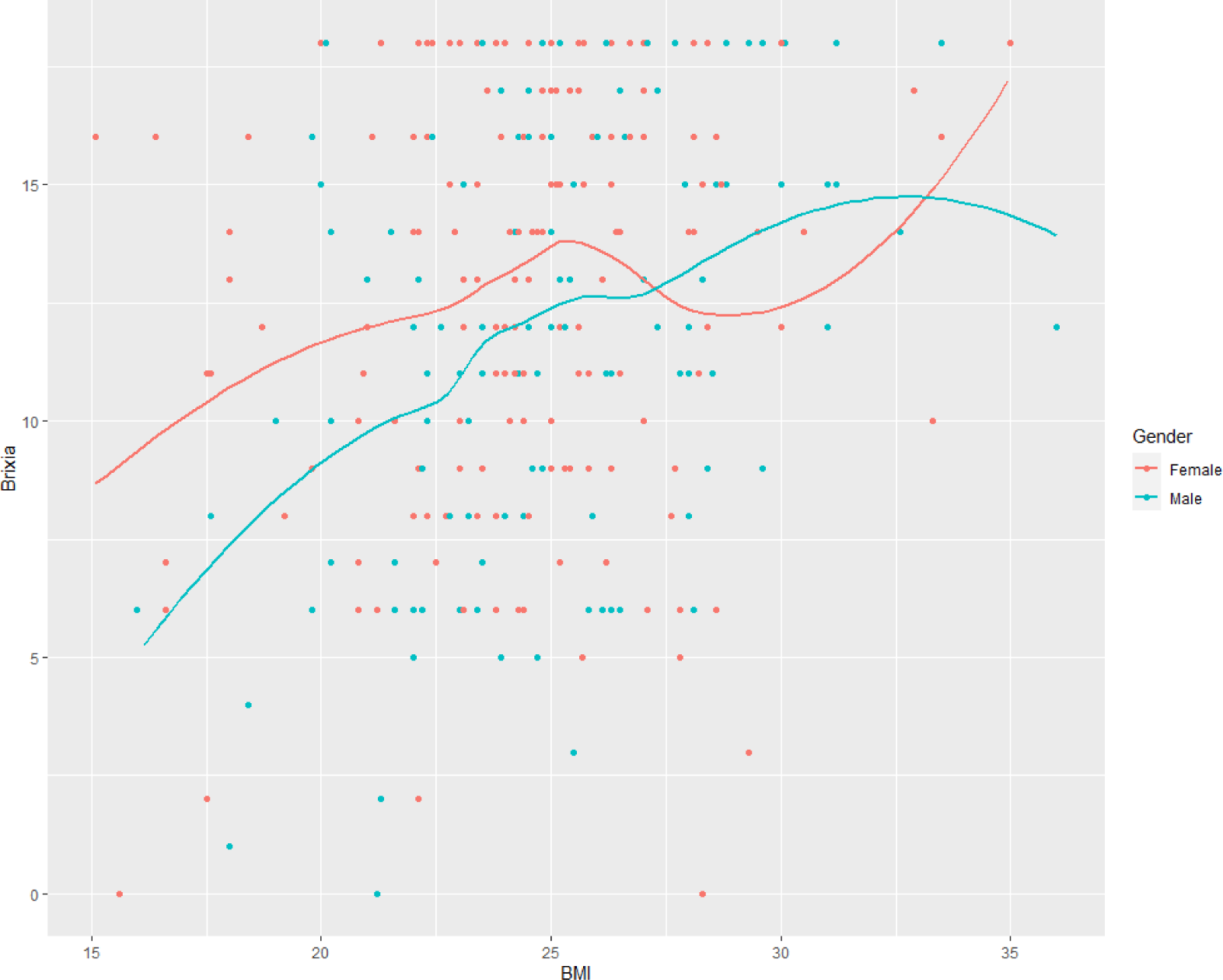
Figure 3 shows the LOESS regression line between the Brixia score and the BMI of men and women. The severity of lung involvement on CXR increased as BMI increased in both genders. Additionally, the Pearson correlation between Brixia score and BMI was 0.25 with a p-value < 0.05. From Figure 4, both men and women in the obese group had the highest Brixia score. Although in the female group, the Brixia score did not change significantly from the underweight group to the obese group (median: 11 to 14). However, in the male group, the median Brixia score ranged from 5 to 13.5.

Table 2 shows the estimate of the coefficient from the Poisson regression for the probability of the Brixia score given the gender and BMI. The estimate of male COVID-19 patients in the obese and underweight groups was significantly different from that of the normal group. There was a dramatic difference in the Brixia scores between men and women statistically. However, we did not find statistically significant differences in the BMI categories of female patients.
4. DISCUSSION
The significance of diagnostic and prognostic approaches in improving patient care for severe COVID-19 was rapidly increasing. The role of CXR was crucial, particularly in circumstances where there was no association between clinical data and respiratory impairment or when CT was unavailable. Many studies have investigated the issue of early prognosis in patients with COVID-19, BMI being one of the most important prognostic factors. Furthermore, various established COVID-19 chest radiographic scoring systems have shown positive results in the prognosis of patients with COVID-19 infection. We conducted this study to demonstrate the correlation between BMI and the severity of pulmonary abnormality, using the Brixia score of hospitalized COVID-19 patients in Vietnam.
In our study, 279 COVID-19 patients had a median age of 59 years (interquartile range 20 to 98 years). The median age was similar to that reported by Sun et al (58.5 with the interquartile range from 47.5 to 69 years. [18]. The median age documented by Au-Yong et al. was higher, at 76 years (interquartile range 59 to 84 years old) [13]. Furthermore, our research reported that the proportion of female hospitalization was greater than that of male patients, with 161 female patients (57.7%) and 118 male patients (42.3%). On the contrary, Malavazos et al. reported that twice as many men as women, with 145 male patients (67%) and 70 female patients (33%) [19]. Similarly, Sun et al. also showed that men were more likely to be admitted than women, with the number of male patients being 72/124 (58%) [18].
The average BMI in our research was 24.6 ± 3.4 kg/m2, and 132 patients (47.3%) and 71 patients (25.5%), respectively, were obese or overweight according to WHO Asian-BMI classification criteria. Malavazos et al., on the other hand, observed a higher mean BMI of 27.5 ± 4.9 kg/m2. According to WHO criteria, overweight and obese patients predominate (overweight 41% and obesity 25%) in this research [19]. Males predominate in the obese category, with 42/54 patients (78%) compared to 12 female patients (22%), and the predominance in the overweight group with 65/87 patients is male (75%). The remaining 22/87 patients were women (25%). Singh et al. reported that hospitalized patients with a fatal result had a mean BMI of 28.53 ± 9.36 kg/m2, and the group that received stable treatment and was discharged from the hospital was 28.74 ± 7.82 kg/m2 [20]. The mechanisms relating obesity and overweight to more severe SARS-CoV-2–related pneumonia remain unclear. First, obesity is known to boost ACE2 expression, which increases SARS-CoV-2 cellular entrance. Secondly, obesity is associated with poor immunological function, which weakens the body and makes it incapable of containing virus reproduction. Thirdly, obesity alters respiratory mechanics, which can influence the mechanisms of lung injury, including atelectrauma, as well as interventions that have been shown to improve the outcomes of ARDS. Finally, obesity has also been associated with the release of inflammatory mediators from adipose tissues, such as tumor necrosis factor and interleukin 6, which can lead to a pro-inflammatory state and oxidative stress, as well as altered mitochondrial bioenergetics in lung epithelial cells, which can lead to an increased risk of acute lung injury [8] [21]. Consequently, having a higher BMI would be associated with a severe course of the disease and hospitalization in COVID-19 patients. Both CXR severity scores and BMI-based obesity are independent predictors of hospitalization and intubation in COVID-19 patients, but obese patients were also at a higher risk of severe CXR than normal-weight patients [9] [16] [21]. Consequently, our additional finding is that lung injury increased proportionally with increased BMI in both sexes, and the obese patient group in both sexes had the highest Brixia score compared to the non-obese patient group. Similarly, in the study by Malavazos et al., obese patients also had a higher median Brixia score (≥ 8 points) [19]. A similar pattern of results was observed in the study by Sun et al., notifying that the obese patient (BMI ≥ 30 kg/m2) were more prevalent in the group with a higher severity score based on chest radiographs. Singh et al. confirmed a similar correlation between BMI and chest radiograph scale, with the chest radiograph score increasing 0.04 units for every 1 unit increase in BMI [20]. These research, however, had been performed using obesity diagnosed by BMI classified by Western standards, depending on the geographic region and racial population.
On the other hands, in this Vietnamese population, if the patient’s BMI, as classified by Asian standards, was higher, the Brixia score for CXR results would be higher. Therefore, in field hospital settings, practitioners can identify high-risk patients based on this aspect for early critical care and prompt transfer to the nearest tertiary hospitals. Furthermore, more research is required, particularly in Vietnamese patients, to verify the utility of this CXR scoring system and its relationship to BMI in predicting the likelihood of the worst outcome. However, in the group of female patients in our study, there was no statistically significant change in Brixia score from the underweight group to the obese group, and vice versa, in the male group, there was a statistically significant change. Although BMI is mostly used to diagnose BMI-based obesity, evaluating fat distribution and abdominal obesity may be a more accurate technique to stratify individuals [21]. Since BMI does not reflect the amount and distribution of body fat, which may differ significantly between people with the same BMI score, it is not consistently the optimal indicator of the obesity [22]. The inconsistent result may be due to discrepancies being attributable to differences in visceral fat deposition between genders. Due to the limitations of clinical work in a confined ward with resource constraints, we were unable to analyze the distribution of fat, but we can assume that the majority of these males were suffering from visceral obesity.
Firstly, our study was a retrospective study based on medical records that would inevitably misrepresent information through the extraction of history and symptoms. Secondly, due to its more complicated indicators and nature to identify COVID-19 pneumonia, Brixia scoring requires the interpretation of a CXR by a skilled radiologist. This could be a problem for primary physicians who need an accurate and simple method to diagnose COVID-19 pneumonia early, but lack the necessary experience and expertise, especially given the shortage of manpower during the COVID-19 pandemic. Finally, age and comorbidities were not included in the model for examining the correlation with BMI due to a lack of sample size. Moreover, we did not find any statistically significant for those variable if we included in our Poisson model.
Conclusion
To our knowledge, this is the first study in Vietnam to reveal a correlation between gender, BMI, and Brixia score on chest radiographs examined by a primary physician in a field hospital. According to the findings, those who had worse pulmonary imaging were mainly middle-aged to elderly, women predominated, and most of them were overweight or obese. The higher the BMI value of the patient, the higher the Brixia score for the findings of CXR which was recorded in both genders, especially for men. We expected that these results of our study will be helpful to clinicians. These data show that for this vulnerable group, prompt access to COVID-19 care, prioritization for COVID-19 vaccination, and other preventive measures need to be taken.


