1. INTRODUCTION
The feet have a high risk of injuries from falling heavy stuff in factories’ environment. In addition, in Viet Nam, we use motorbikes as a predominant vehicle in traffic. Thus when a vehicle accident occurs, the feet are often injured because of rubbing with the road surface. The feet have thin skin, and less subcutaneous tissue than other parts of the body. Hence feet injuries often include loss of skin, and exposure of tendon, bone, vessel or nerve. Without good strategy treatment, exposing tissues will be necrotic resulting in reduced movement function of the feet, effected to everyday life and working effectiveness of patients, so we need to give an effective solution for covering skin defects in the foot.
With respect to cover skin defects in the forefoot region, Liu et al. suggested using the intermediate dorsal neurocutaneous flap, but it serves to small size defects (1). The modest defects are often covered by pedicle flaps. The most popular is the sural neurocutaneous flap (2, 3). However, the sural flap is predominant to resurface heel defects and hints foot. When using the sural flap on the forefoot has, there are some disadvantages, such as being thick and bulky and effected to the aesthetic result on the ankle and foot (4). The alternative choice is a lateral supramalleolar flap nourishing by the dorsal vessel anastomosis with the peroneal artery (5). This flap has the same advantages as the sural flap, such as not sacrificing the main artery of the lower limb, the long pedicle, and it is used more popular recently (6, 7).
In Viet Nam, there are few case reports about the effectiveness of the lateral supramalleolar flap nourishing by the dorsal vessel anastomosis with the peroneal artery, with good results, but the elevated flap technique was a tough challenge to surgeons. At the Hospital for Traumatology and Orthopaedic, Ho Chi Minh City, we have used this technique for covering skin defects in the foot with a good outcome. This study aims to clarify the effectiveness of this flap. The result from this research can help inexperienced doctors to have more clinical evidence and feel more confident in using this type of technique.
2. MATERIALS AND METHOD
This prospective observational research was run from December 2017 to December 2021 at the Hospital for Traumatology and Orthopaedics, Ho Chi Minh City.
During the research, we gathered information from 27 cases treated with lateral supramalleolar flap nourishing by the dorsal vessel anastomosis with the peroneal artery surgery for covering skin defects in the subunits of the foot. The follow-up time was at least six months and a maximum of 2 years. Included criteria were cases with foot skin loss and exposed important structures such as bone, nerve, tendon and vessels. Excluded criteria were patients with crush injuries in the lower part of a leg, diabetes and peripheral vascular disease.
Ethical approval was permitted by the University of Medicine and Pharmacy at Ho Chi Minh City (280/HÐÐÐ-ÐHYD) and was in line with the Helsinki Declaration. The participants and their caretakers were provided the information and the purpose of the study, then they were requested to sign a written informed consent form and permission to publish their pictures and information in this research (8).
In the research, we gathered demographic information, etiology, size of skin defect, size of flap, time of surgery, and post operation data. The success criteria were covering a skin defect in one procedure by the flap. Failure criteria were occurring post operation flap necrosis which needed a skin graft later. The criteria used to define survival of the flap were: the flap was fully alive, and the wound was healed at the first stage. Distal part necrosis was defined as necrosis of less than 40% of the total flap area in the distal site. Half of flap necrosis was the necrosis of 40% to 70% of the whole flap area. Total flap necrosis was the necrosis of more than 70% of the entire flap area.
We measured the size of the defect first and later we drew a sample figure of the defect on the lateral border of the leg by adding 0,5 cm to provide flap shrinking after dissection. Flap designed with the subcutaneous pedicle as Valenti suggested, the width of the pedicle was estimated to be at least 3cm (9). We identified a pivot point by an intersection of the line connecting two malleoli and extending the fourth phalange axial (8). We incised the anterior edge to the deep fascia of the flap, and we observed the emerging point of the perforator branch of the peroneal artery on the interosseous membrane first. After that, we dissected the descending branch up to the pivot point. We tried to get superficial veins as much as possible in the subcutaneous pedicle. We also used the modified technique to dissect the skin pedicle with the width of subcutaneous tissue 3cm and 1cm skin on the middle of the pedicle. This modification was used when the under-skin tunnel was incised to avoid the stress on the pedicle with advantages such as preventing skin graft on the subcutaneous pedicle and dog ear effect at the pivot point. At this point of flap elevation progress, we tied and separated the emerging perforator branch and incised the flap’s border as drew the figure. We shifted the dissected flap to cover the injured site. We closed the donor site with a split skin graft.
We used frequency and percentages to describe qualitative data, such as the location of a defect, causing of an injury, a flap outcome, a leg site, and associated injuries. Along with quantitative statistics such as a size of an injury, a size of a flap, age, and time of surgery, we used mean or median when appropriate. All statistic analyses were run using Stata, version 16 (8).
3. RESULTS
The follow up period in our study ranged from 6 to 24 months with a mean follow up of 11,3 months. There were 27 patients with foot defects that were covered by lateral supramalleolar flap nourishing by the dorsal vessels anastomosis with the peroneal artery (Table 1). Two-thirds of cases were male, and the average age in the study was 42 (ranging from 15 to 68 years old). The predominant cause of injury was a traffic accident (81%). Alternative sources were labor accidents, burns, and chronic ulcers. The mean size of the defect was 36 cm2 (ranging from 9 cm2 to 80 cm2). The average size of the flap was 46 cm2 (ranging from 12 cm2 to 104 cm2). The mean surgery duration was 92 minutes (ranging from 60 - 125 minutes).
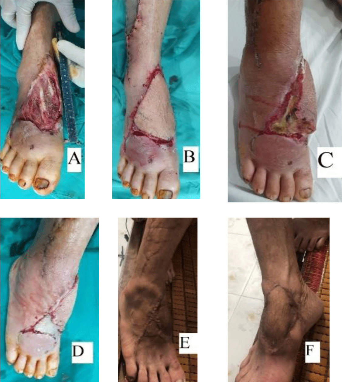
In our study, there were three partial necrosis flaps that later needed debridement and a second surgery to solve. The first partial flap belonged to 61 years old patient with a 6cm × 11cm wound at the middle foot. We elevated a 7cm × 12cm flap with the subcutaneous pedicle tunneled under the skin. After 1 week post operation, the flap necrotized partially. We performed a second surgery to debride and spit skin graft to cover the wound (Figure 1). The second case was 50 years old with a 6cm × 12cm defect at the middle extending to the forefoot, used a 6cm × 11cm size of the skin flap, and the roof of the skin tunnel was incised to set in the pedicle. After one week, the flap necrotized at the distal part and was solved by debridement with a split skin graft. The third case was 51 years old with a 5cm × 11cm skin defect at the middle extending to the forefoot. We covered it with a 5.5cm × 12 cm skin flap with the pedicle set in the incised bridge skin. The flap was gradual necrosis at the distal part and was fixed by a split skin graft in the second surgery. Particularly, there was one case in our study with a distal part of flap necrosis, but it simply needed a minor surgery debridement and suturing. The wound later healed uneventfully (Table 3).

| Variables | Mean | Minimum | Maximum |
|---|---|---|---|
| Age (years) | 42 | 15 | 68 |
| Size of the defect (cm2) | 35,6 | 9 | 80 |
| Size of the flap (cm2) | 46 | 12 | 104 |
| Intraprocedural time (Minutes) | 92 | 60 | 125 |
| Alternative intervention | Flap outcome | ||
|---|---|---|---|
| Half of flap necrosis | Distal part necrosis | 100% alive | |
| Minor surgery | 0 | 1 (4%) | 0 |
| Skin graft | 3 (11%) | 0 | 0 |
| None | 0 | 0 | 23 (85%) |
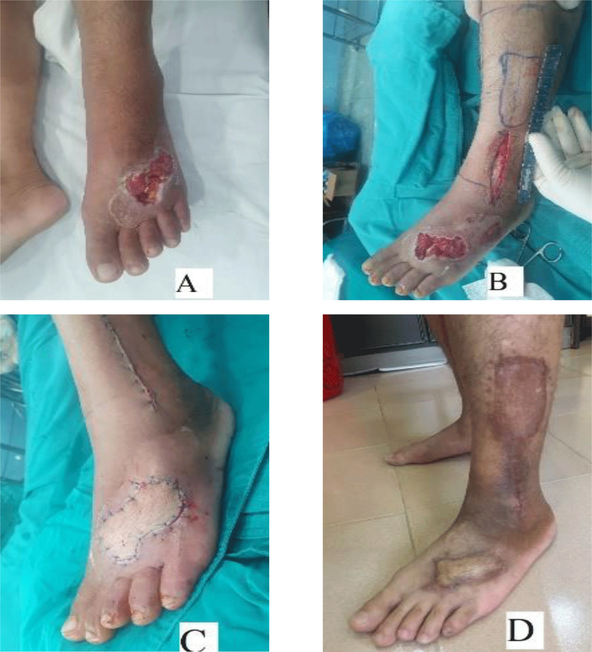
The success rate in our study was 89% (n= 24/27) (Table 4). The long term outcome showed that almost all patients had flap color the same as the dorsal foot. The flap thickening was higher than the vicinity skin, but it did not affect wearing shoes. The donor site was covered by a split skin graft with a 100% success rate. There was not any case with an adhesion tendon in our study (Figure 2).

4. DISCUSSION
In 1988, Masquelet et al. suggested using a lateral supramalleolar flap, which was dissected from the lateral lower site of the leg to cover the defects in the ankle and the foot (5). In 1991, Valenti modified this elevating technique of this flap, thoroughly enhance the length of the skin flap by including a long subcutaneous pedicle (9). Furthermore, some anatomy of blood supply of this flap research were reported, strengthening the usefulness of this flap (10-12). However, the authors reported the results from the small size study of using lateral supramalleolar flap nourishing by the dorsal vessels anastomosis with the peroneal artery such as Voche reported eight flaps in his research (6), Nambi reported three flaps in 2020 to cover the defects in the hindfoot (7). In our study, 27 flaps were used with an encouraging success rate. In the literature, authors showed that elevating this flap was a challenging technique. The surgeons must meticulously isolate the emergence perforating branch of the peroneal artery, later dissecting the descending branch up to the anastomosis point with dorsal vessels. These branch vessels are located sub fascia deeply, under tendons, and above the ankle capsular ligament. Thus, surgeons need gentle manipulation to avoid vessel injuries iatrogenesis.
The lateral supramalleolar flap nourishing by dorsal vessels anastomosis with the peroneal artery can be dissected as a skin flap with significant size designing, and the donor site is healed eventually by a split skin graft. In our study, the biggest size of the skin flap was 104 cm2 (Table 2). The injuries' location were almost in an awkward position, such as in the middle extending to the forefoot (Table 4). This could be an indication that the potential using of the lateral supramalleolar skin flap is significant to cover the defects in problematic areas in the foot. Some authors suggested to use the modify of later supramalleolar flap such as a subcutaneous flap. After transferring the subcutaneous flap to cover the defect, the flap’s surface was covered by the skin graft, and the initial result was good (13, 14). However, this modified method could not use to cover large defects instead of small and median defects. In addition, it was hard to follow the vital sign of the subcutaneous flap because of the skin graft on the flap’s surface.
The surgery time of the lateral supramalleolar flap was short. In this research, the mean surgery time was 92 minutes (Table 2). This period is faster than the surgery time of the free flap (15). The short intraprocedural time requires less procedural sedation. Thus it decreases the complication risks from sedation medicine. Additionally, Hang Cheng et al. reported that prolonged operative time could increase the chance of surgical site infection (16). Besides, free flap surgery usually requires two teams of surgeons such as one team for dissecting the flap and another for preparing the vessels at the recipient site. Compared to lateral supramalleolar flap surgery just needs one surgeon and an assistant surgeon to perform.
Regardless of the high success rate which showed in our research (89%), some lessons can be achieved from the failure cases. For example, the first failure case had a subcutaneous pedicle. We realized that this case had significant skin flap size. Thus tunneling the pedicle reduced the venous outflow that led to flap congestion which resulted in partial necrosis of the skin flap. We suggest that the surgeons consider more before deciding to use the tunneling pedicle technique to ensure the flap’s vital. With another two failure cases, they had large skin defects. The skin flaps were elevated by a significant size. Although the pedicles were set in an incised ceiling skin to reduce the stress on them, the flaps still congested and later necrotized the distal part. Some authors suggested using local subcutaneous heparin injection to save congestion flaps in the postoperative observation period (17). However, we have not experienced this method used practically.
In our study, we had a limitation that the research was performed on a small size of cases, particularly in the children group. The leading reason was the entanglement of dissecting approach due to the very small pedicle vessel in this group. However, in our study, a fifteen years old patient was covered by this flap with success result. This result showed the potential usefulness of this covering technique for children patients when it is performed by experienced surgeons.
Conclusion
The present study showed that the initial results of applying the lateral supramalleolar flap nourishing by the dorsal vessels anastomosis with the peroneal artery for covering skin defects in sub-units of the foot with high success rates, the significant size of flap designing and shorter intraprocedural time comparing to free flap technique.


