


1. INTRODUCTION
Melioidosis was first described as a “Glanders-like” disease in 1911 by Whitmore and Krishnaswami, caused by the obligatory aerobic gram-negative bacillus B. pseudomallei [1]. From then, it emerged as a sporadic but fatal infectious disease in South and Southeast Asia, Northern Australia and China. The bacterium can be found in wet soil and surface water, infiltrating to the host through skin abrasion, digestive or respiratory tract. Up to 60% melioidosis patients have diabetes mellitus and other risk factors related to immunocompromised conditions such as cirrhosis, chronic renal disease, chronic lung disease, malignancy, long-term corticosteroid therapy, alcoholism. Melioidosis presents a variety of clinical symptoms from asymptomatic carriage to pneumonia (most common), skin and soft tissue infection, body organ abscess, encephalomyelitis, genitourinary infection and uncommon bone and joint infection [2]. Since the first case reported in 1925, Vietnam have only recorded about a hundred cases over a century, most of whom manifested with septicemia and pneumonia and had high mortality rate [3].
Septic arthritis is a common orthopedic emergency infection, typically presenting with swelling, erythema, tenderness, and limited range of motion of the affected synovial joint. The primary treatment requires early removal of pus and antibiotic administration, without which the condition may lead to irreversible joint destruction and even death. Staphylococcus aureus and Streptococcus species stand as the most prevalent causative gram-positive bacteria globally, meanwhile gram-negative bacteria such as Escherichia coli and Pseudomonas species are also frequently observed in elderly patients with concomitant diseases [4]. Septic arthritis caused by B. pseudomallei shares similar signs and symptoms to the above causative agents but requires a distinct antibiotic regimen, consisting of an intensive phase followed by a prolonged eradication phase, to prevent mortality and relapse. In addition, this condition has been modestly documented with scattered case reports from around the world [5]. We hereby report such a case with two consecutive incidents of septic arthritis that we had not encountered in clinical practice or found in any domestic publication.
2. CASE PRESENTATION
A 60 year-old male living in Tay Ninh province presented to our hospital with acute pain in his left hip and fever of a 5-day duration. He was a farmer with a 4-year history of type 2 diabetes mellitus, gastroesophageal reflux disease and Crohn’s disease which was being managed with 8 mg methylprednisolone and 2,000 mg mesalazin daily. His fever was intermittent and accompanied by rigors and chills. The left hip pain was insidious at the onset, but gradually worsened that made him nearly bedridden and restrict any joint movement. He reported no recent trauma or other complaints.
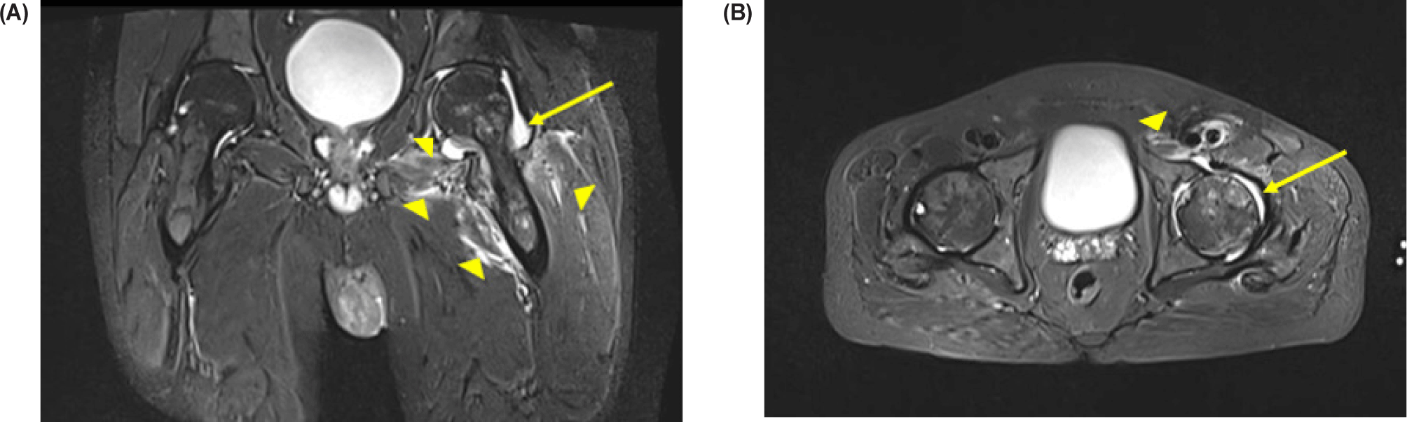
On examination, he was pale with a body temperature of 38.5°C. There was no apparent swelling, warmth, or redness in the left hip area, but there was tenderness in front of the groin and he experienced severe pain with passive hip motion in any direction. Initial laboratory tests revealed his peripheral white blood cell count (WBC) of 16.2×10⁹ /L with 82.6% of neutrophils, hemoglobin level of 106 g/L, serum creatinine level of 1.18 mg/dL, C-reactive protein (CRP) level of 222 mg/dL, hemoglobin A1C level of 9.1%. The chest and pelvic X-rays were normal but the ultrasound detected a thin layer of fluid within the hip capsule. Because of high suspicion of joint infection, an magnetic resonance imaging (MRI) with intravenous gadolinium contrast was carried out which showed effusion in the left hip joint with the maximum fluid layer thickness of 10 mm, increased signal from the joint capsule and the surrounding muscles (Fig. 1).
Blood and synovial fluid samples were collected for culture and antibiogram. However, we only aspirated a small amount of yellowish fluid under ultrasound guidance due to the thin layer of intracapsular effusion. Afterward, the arthrocentesis needle and syringe were rinsed with 2 mL of normal saline, and the specimen was sent to the Microbiology department.
After the sample was obtained, the patient was administered the empirical antibiotic therapy with intravenous ertapenem and vancomycin that provided broad-spectrum antimicrobial coverage while awaiting the culture results. After 48 hours, he experienced partial relief from hip pain, but still had a high-grade fever, while the inflammatory markers slowly decreased with WBC of 13.6 ×10⁹ /L, CRP of 167.6 mg/L. The culture reports came after 3 days. It showed growth of B. pseudomallei in both the blood and synovial fluid samples with sensitivity to amoxicillin/clavulanic acid, trimethoprim/sulfamethoxazole, ceftazidime, imipenem and meropenem. Definitive therapy was started with intravenous meropenem 1,000 mg every 8 hrs and oral co-trimoxazole 1,600/320 mg every 12 hrs (intensive phase). The patient quickly responded to this regimen. In addition, blood sugar levels and Crohn’s disease were managed, and 5 mg oral folic acid was also administered to prevent antifolate activity of co-trimoxazole. Physiotherapy was employed to improve joint mobilization without any surgical drainage necessary. For personal reasons, he decided to continue the intensive phase of intravenous antibiotic at his provincial hospital after 2 weeks of treatment, when he was completely free of fever and hip joint pain with WBC of 7.47×10⁹ /L, CRP of 5.3 mg/L, creatinine of 0.92 mg/dL and a negative second blood culture. He was transferred with a detailed laboratory and treatment report, accompanied by a clear plan to complete the intensive phase as well as an examination appointment at our outpatient clinic.
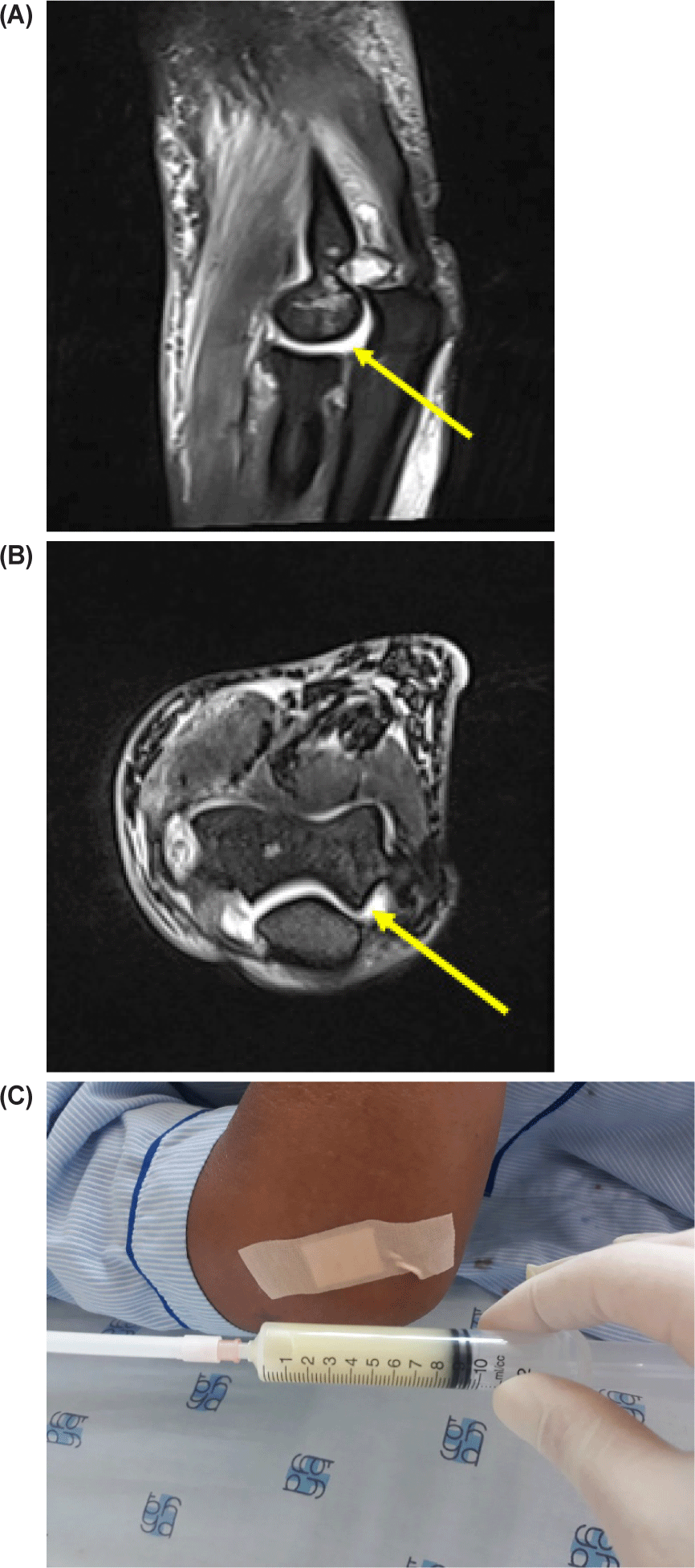
Unfortunately, he was only given an additional week of intravenous antibiotic before being sent home with oral ciprofloxacin as the sole treatment. A month later, he returned to our department with noticeable swelling, warmth, redness, and tenderness in his right elbow, coupled with significant limitation in range of motion. Laboratory tests showed WBC of 21.28×10⁹ /L, CRP of 108 mg/L. Radiography indicated a normal X-ray of the elbow but a large joint effusion with increasing signal intensity of surrounding tissue on MRI. Blood culture was negative but joint aspiration at ward revealed purulent fluid with cloudy yellow color, which was positive for the same microbial agent and had similar antibiotic sensitivity as the first time (Figs. 2–4).
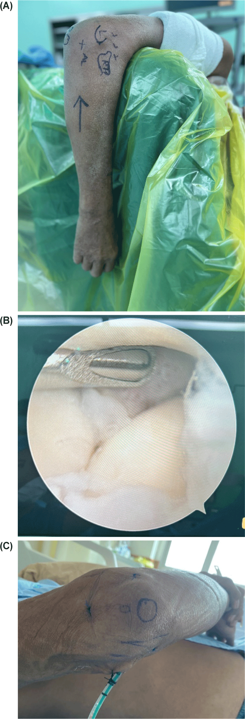
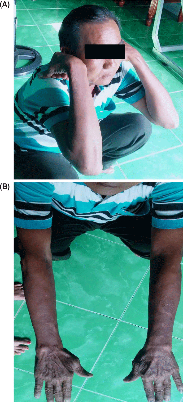
We urgently performed an elbow arthroscopy for pus drainage and debridement and restarted a 4-week intensive antibiotic phase, and prescribed a 6-month outpatient eradication phase with oral co-trimoxazole 1,600/320 mg every 12 hrs. Once again, the patient recovered well and regained full range of motion and function of his elbow. He continues management for diabetes mellitus and Crohn’s disease at our hospital without any infectious relapse.
3. DISCUSSION
Melioidosis was previously considered rare; however, the number of confirmed cases is rising each year due to improvements in awareness and recognition in Vietnam [3]. A significant number of French and American soldiers contracted the disease during the Vietnam war [6],[7], highlighting that the country lies within the endemic region with a high bacterial reservoir in soil and water resources. Our patient came from a rural area where he was frequently exposed to the field environment. Additionally, diabetes mellitus and long-term corticosteroid and mesalazin use for Crohn’s disease put him at a heightened risk of infection.
Despite being a well-recognized manifestation of melioidosis, septic arthritis is rare, particularly when it occurs in isolation without other organs involvement. In a study conducted in northern Australia involving 540 patients, only 4 cases (0.7%) were diagnosed with septic arthritis [8]. In a high-endemic region like Thailand, the number was slightly higher with 81 cases (11.9%) out of 679 patients in a retrospective study by P.Teparrakkul et al [9]. However, many of these cases were associated with other foci, and only 20 patients had involvement of more than one joint. Among joints, knee was the most commonly involvement, followed by ankle, hip and shoulder. Elbow and other small joints were very rarely affected [9],[10]. This is the first time we recorded an isolated consecutive melioidosis septic arthritis case with hip and elbow involved.
It was clear that our patient acquired joint infections through the bloodstream, evidenced by a positive bacteremia upon the first hospitalization, without any identifiable entry point. In the second admission, the negative blood culture could be due to prior antibiotic use. Hematogenous spread is also the most common way of transmission in melioidosis bone and joint infection [5]. The bacterium can also directly spread from adjacent infectious organs or soft tissue.
B. pseudomallei infection does not have specific clinical features and can be challenging to distinguish from other causative agents such as Staphylococcus, Streptococcus or other purulent bacteria. Patients typically have fever, pain, and limited mobilization of the affected joint. Examination usually shows a swelling, redness, warmth, and tenderness joint, accompanied by elevated WBC, CRP, procalcitonin and erythrocyte sedimentation rate. Due to its uncommon occurence and symptom similarity with other causative agents, melioidosis septic arthritis can often go unrecognized and misdiagnosed [5]. Therefore, collecting blood and synovial fluid samples for culture before initiating empirical antibiotic treatment is extremely important and can be seen as the gold standard in the diagnosis. In our case, we had to rinse the hip arthrocentesis needle and syringe with normal saline to obtain the required amount of fluid as requested by the Microbiology department. The positive result demonstrates that a very small sample can hold significant importance and should not be discarded. Serological antibody tests such as indirect hemagglutination and immunofluorescence assay can be useful in cases of negative culture but there is still strong suspicion of infection. However, their specificity is low and should be interpreted cautiously in endemic areas, where even healthy people can raise the antibody due to repeated exposure to this organism during outdoor activities [11],[12]. Molecular tests such as PCR or pulsed-field gel electrophoresis are also utilized in certain laboratories, although they are generally less sensitive than the culture method [13].
Prompt antibiotic administration and surgical intervention for pus drainage and debridement are the cornerstone of melioidosis septic arthritis treatment. There is a high consensus about antibiotic regimen which is divided into 2 phases. The intensive phase is recommended with intravenous ceftazidime or meropenem or imipenem for a minimum duration of 4 weeks, combined with oral trimethoprim+sulfamethoxazole as an adjunctive therapy due to its excellent tissue penetration. The eradication phase begins immediately following initial intensive phase to prevent relapse. Oral trimethoprim+sulfamethoxazole is the first choice and should be maintained for at least 3 months [5],[14]. Septic arthritis is an indication of surgical intervention, and in this particular case, we had devised a plan for hip debridement already. However, a careful consideration is necessary because the hip joint is situated deep and surrounded by substantial tissue, making the approach become challenging. Even with the use of an arthroscope, the procedure requires an experienced surgical team and carries a high risk of infection spread due to fluid pumping. This patient had a small amount of hip joint fluid and responded remarkably well to antibiotic treatment, leading us to decide against surgery. There are also some case reports of melioidosis septic arthritis successfully treated with antibiotic alone, which strongly support our decision [15],[16]. In contrast, the elbow joint is much easier to approach; and an large pus effusion with severe tenderness and extremely high inflammatory index made us decide an urgent operation in this case to preserve the cartilage as well as reduce the bacterial load. Arthroscopic debridement is the fisrt-choice procedure for treating septic arthritis due to its minimal invasive, allowing effective joint evaluation, synovectomy and lavage, which provides better outcomes compared to open debridement in both infectious eradication and joint function [17]. This decision also resulted in a favorable post-operative outcome in this case.
Melioidosis can have the potentiality of latent infection or relapse, sometimes after years, which has given the condition the nickname “Vietnam time bomb” [3]. A high relapse rate (up to 23%) was reported in patients, especially who received antibiotic regimen less than recommended time, severe disease with multiple foci and positive blood culture, poor compliance [18]. Relapse should be managed in the same way as the first episode [18], as was applied in our case. The patient is still being in close follow-up for any sign of recurrence.
4. CONCLUSION
Melioidosis septic arthritis is rare, but should be considered in patients at high risk, especially those from endemic areas. Blood and synovial fluid cultures are the gold standard for diagnosis. The treatment includes prompt parenteral antibiotic, either combined with surgical debridement, depending on the circumstance. Regardless of the method, long-term eradication antibiotic therapy is crucial to prevent relapse and the development of resistant organisms.