case report

Extrapulmonary Intrapleural Hydatid Disease- A Case Report

Muhammad Usman Hashmia,*, Muhammad Kaleem Ullahb, Mohsin Sarwara, Alia Sultana, Abdul Aleemb, Iftikhar H. Khanb
Author Information & Copyright
aShifa College of Medicine Islamabad, Pakistan
bNishtar Medical University Multan, Pakistan
*Address correspondence: Muhammad Usman Hashmi at Shifa College of Medicine Islamabad, Pakistan; Tel/Fax: +92-333-598-9487; E-mails: muh.haashmi@gmail.com

© Copyright 2019 MedPharmRes. This is an Open-Access article distributed under the terms of the Creative Commons Attribution Non-Commercial License (http://creativecommons.org/licenses/by-nc/4.0/) which permits unrestricted non-commercial use, distribution, and reproduction in any medium, provided the original work is properly cited.

Received: Jan 12, 2019; Revised: Jan 29, 2019; Accepted: Feb 19, 2019

Published Online: May 31, 2019

Abstract

Hydatidosis is a zoonotic disease caused by the tapeworm Echinococcus granulosus. It mostly involves liver and lungs whereas the unusual sites are spleen, kidney, peritoneal cavity, skin, muscles, heart, brain, vertebral column, ovaries, pancreas, gallbladder, thyroid gland, breast, and bones. Herein, we describe a unique case of intrapleural hydatid cyst which was solely confined to the pleural space without involvement of lung parenchyma and other associated structures. Despite the fact that Pakistan is an endemic country for hydatid disease, we could not identify any single documented case of intrapleural hydatid disease from Pakistan. The purpose of our case report is to promote awareness among fellow healthcare professionals about this rare variant of hydatid disease and to prevent any missed diagnosis and life-threatening complications. Hence, once the diagnosis was established, we performed a right-sided thoracotomy. Intraoperatively, the lung was collapsed due to the pressure effects of hydatid cysts. The hydatid cysts were evacuated completely from the pleural cavity and postoperatively the patient had an uneventful recovery and remained asymptomatic during all the follow-up visits. Conclusively, the hydatid disease may involve unusual sites such as the pleural space. A strong clinical suspicion, supportive radiological findings, and positive serological evidence play a critical role in the establishment of the diagnosis.

Keywords: Hydatid disease; echinococcus granulosus; intrapleural; extrapulmonary; case report

1. BACKROUND

Human hydatid disease or cystic echinococcus is a parasitic infestation caused by the larval form of the tapeworm Echinococcus granulosus, which lives in intestines of dogs and other canines. Ingestion of feces contaminated with parasite eggs causes liberation of larvae in the duodenum of intermediate hosts which can be sheep, goat, reindeer or human. The analysis of recent medical literature reveals that the incidence of hydatid disease has increased worldwide especially in tropical countries [1]. Therefore, the World Health Organization (WHO) has declared it as the most neglected tropical disease [2]. This zoonotic disease is more prevalent in sheep-raising areas like the Mediterranean countries, South America, northern China, the Middle East and India [3]. The liver is the most commonly involved site (75%), followed by the lungs (15%), spleen (5%), and other organs (5%) [3]. The authors present a case of intrapleural hydatid cyst which was solely confined to the pleural space sparing the lung parenchyma and other associated structures.

2. CASE PRESENTATION

A 10-year-old male presented to the local basic health unit of Dera Ismail Khan, Pakistan. His chief complaints were exacerbating severe chest pain associated with high-grade fever and dyspnea. In addition, he complained of shortness of breath, dry cough and vague chest pain that developed over the last three months. Initially, his symptoms were mild but progressively worsened over time. The patient also reported appetite loss, without any other complaint such as palpitations, nausea, jaundice, or vomiting. There was no history of change in bowel habits or urinary symptoms. The patient was living in a rural area and he had a history of long-term exposure to domestic pets. He did not undergo any kind of surgery in the past.

At the time of presentation, the patient was hemodynamically unstable with a pulse rate of 108 beats per minute, blood pressure of 100/80 mmHg and an oral temperature of 39°C. The patient was tachypnic with a respiratory rate of 26 breaths per minute. Chest movements and breath sounds were decreased on the right side with a dull percussion present in comparison to left hemithorax examination. There were no additional breath sounds. Chest X-ray was ordered which revealed right-sided lung white-out with contralateral mediastinal shift. The physician at the local health facility diagnosed it as pyothorax and placed a chest drainage tube on the right side. Nevertheless, two weeks after the placement of right-sided tube thoracostomy the condition of the patient did not improve. Subsequently, he was referred to the Department of Thoracic Surgery, Nishter Medical University Hospital, Multan Pakistan, for further evaluation and management. The patient was thoroughly re-assessed. Table 1 describes the laboratory results of baseline investigations.

Table 1: Summary of the laboratory data on admission
Initial investigations Value Reference range
Complete blood count
White blood cells 15,600/μL 4500-11,000/μL
Hemoglobin 13.2 g/dL 13.1–17.6 g/dL
Hematocrit 42.1% 38.1-50.8%
Mean cell volume 87.6 fL 84.6-100.6 fL
Platelet counts 176,000/μL 150,000-400,000/μL
Coagulation
PT-INR 1.1 0.89-1.12
aPTT 36 seconds 25-40 seconds
Biochemistry
Total bilirubin 0.89 mg/dL 0.1–1.2 mg/dL
Aspartate aminotransferase 27 IU/L 12-35 IU/L
Alanine aminotransferase 24 IU/L 6–40 IU/L
Lactate dehydrogenase 197 IU/L 119-229 IU/L
y-Glutamyl transpeptidase 31 IU/L 0-48 IU/L
Blood urea nitrogen 10.6 mg/dL 7.4-19.5 mg/dL
Creatinine 0.91 mg/dL 0.5-1.2 mg/dL
Total protein 6.7 g/dL 6.4-8.3 g/dL
Albumin 3.7 g/dL 3.8-5.2 g/dL
Serum Sodium 134 mEq/L 135-147 mEq/L
Serum Potassium 4.1 mEq/L 3.4-4.8 mEq/L
Serum Chloride 103 mEq/L 98-110 mEq/L
Glucose 69 mg/dL 70-110 mg/dL
C-reactive protein 1.68 mg/dL 0-0.5 mg/dL
Anti-Hepatitis C antibodies by ELISA Negative
HBsAg by ELISA Negative

APTT: Activated partial thromboplastin time;

PT-INR: Prothrombin time international normalized ratio;

ELISA: Enzyme-Linked Immunosorbent Assay:

HBsAg: Hepatitis B surface antigen.

Download Excel Table

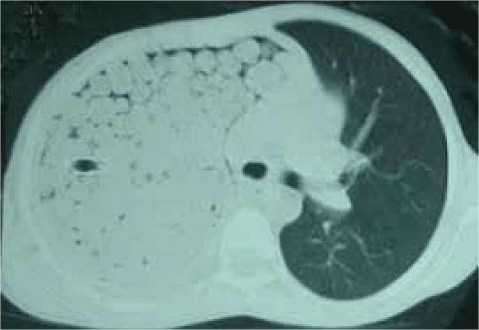
A chest CT scan was also ordered, showing multiple cysts occupying the whole intrapleural space on the right side and completely compressing the right lung. Fig. 1 shows the chest CT scan of the patient revealing a large mother hydatid cyst.
mpr-3-1-12-g1
Figure 1: Chest CT scan of the patient showing multiple cysts that filled the whole pleural space on Right side
Download Original Figure

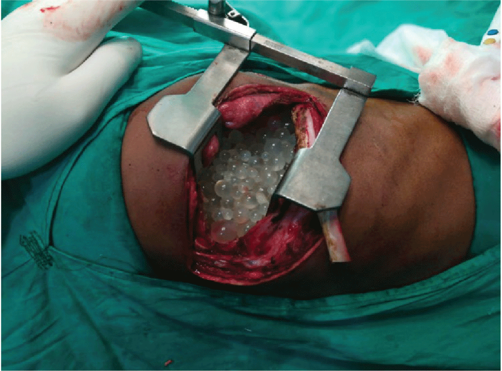
The mother hydatid cyst also contained numerous daughter cysts of variable size occupying the whole pleural space on right side. Based on chest X-ray and CT scan findings, a suspicion of intrapleural hydatid disease was made. The patient underwent right posterolateral thoracotomy. The right-sided pleural cavity was obliterated by a large mother hydatid cyst containing multiple daughter hydatid cysts. In addition, the right lung was totally collapsed due to the compressive effect applied by the multilocular cystic mass. Intraoperatively, the evacuation of cysts content resulted in immediate lung expansion and no other abnormality or defect was identified in the lung parenchyma. Fig. 2 depicts the intra-operative findings of this case where numerous daughter cysts are rushing out from the thoracotomy site.
mpr-3-1-12-g2
Figure 2: Intra-operative findings of the case. Numerous daughter cysts are rushing out from the thoracotomy site.
Download Original Figure

The wound and pleural cavity were irrigated with normal saline and the collapsed lung was adequately infl via the assistance of positive pressure ventilation. The patient had an uneventful recovery and was discharged from the hospital three days postoperatively. The patient did not have any air leakage through the chest tubes during his post-operative course, confi the non-involvement of lung parenchyma. He was consequently followed up at the intervals of one week, one month and six months. During all these visits the patient remained asymptomatic and chest X-ray performed at the follow-up visits showed bilaterally normal lungs. All these events are summarized in table 2 in their chronological order.

Table 2. Timeline for the case report showing chronology of the various events
Dates Relevant Past Medical History and Interventions
The resident of a rural area. A positive history of long-term exposure to domestic pets. No record of vaccination. No significant past medical history. Never underwent any kind of surgery in the past.
Date Summaries from Initial and Follow-up Visits Diagnostic Testing Interventions
30-01-2018 First visit at basic health Unit of Dera Ismail Khan
Chief Complaints:
Exacerbation of chest pain with worsening dyspnea.
The patient also complained of fever.
Physical Examination:
A hemodynamically unstable patient with decreased chest movements and breath sounds on the right side with a dull percussion note in comparison to left hemithorax examination. There were no additional sounds.
Diagnosis:
Right-sided pyothorax
Plain chest X-ray revealed white out of the right lung. Right-sided tube thoracostomy
05-02-2018 Follow-up at basic health unit Dera Ismail Khan: No improvement, and symptoms worsening over time. The output of the chest drainage unit was nil with absent air leakage.
The patient was referred to Nishtar Medical University Hospital Multan for further evaluation and management.
Repeated chest X-ray showed similar findings.
06-02-2018 The patient was admitted to the Thoracic Surgery Department of Nishtar Medical University Multan, in order to be re-evaluated and furtherly assessed. Subsequently, an extensive workup revealed the diagnosis of hydatid disease. He was operated one day later and had an uneventful recovery. He was discharged on the third postoperative day. Baseline laboratory investigations were performed. Chest CT scan was also ordered which showed a large hydatid cyst with multiple daughter cysts. Anti-hydatid IgG antibodies also came positive Right-sided thoracotomy for evacuation of multilocular hydatid cystic mass.
Chest physiotherapy and incentive spirometry were advised.
17-02-2018 First follow-up visit after surgery. The patient was asymptomatic with no air and fluid leak from the chest drainage unit. Plain chest radiograph revealed expanded lung fields with a minor pleural effusion. Repeated chest X-ray
19-03-2018 Second follow-up visit after surgery. The patient remained asymptomatic with fully expanded lung fields and resolution of pleural effusion. Chest X-ray
20-09-2018 Third follow-up visit. The patient was asymptomatic. There was no recurrence of the disease. Chest X-ray
Download Excel Table

3. DISCUSSION

Hydatid disease has become an increasingly emerging public health problem in Pakistan. However, the medical literature from Pakistan is quite scant and does not address this issue adequately. A recent study by Ahmad et al claims that between 1980 and 2015, only three research papers on the prevalence of hydatid disease and 13 case reports about the involvement of unusual body locations by hydatid cysts were published [4]. The clinical presentation of disease correlates with the involvement of particular organs and adjacent structures. Pezeshki et al stated the relative frequency of different unusual sites of hydatid cysts such as spleen (7.69 %), abdomen (3.84 %), spinal cord (2.56 %), sub-diaphragmatic space (1.28 %), peritoneum (1.28 %), kidney (1.28 %) and pancreas (1.28 %) [5]. The extra-pulmonary, intrapleural hydatid disease is a rare clinical entity even in endemic countries, though the pleural cavity is not completely immune to Hydatid disease. Thameur H, et al. in their review of 1,619 cases, found 42 cases of intrathoracic extra-pulmonary hydatid disease. Furthermore, out of the aforementioned 42 cases, only 22 were primary intra-pleural hydatid cysts [6]. An Indian study by Srinivasan B et al reports that out of 127 cases only 4 were extrapulmonary and intrapleural [7]. Nevertheless, we could not identify any study from Pakistan reporting extra-pulmonary intrapleural hydatid cysts sparing the lung parenchyma. It is important to note that in the case of primary pleural hydatid cyst disease, the larvae infest the pleura either through hematogenous or lymphogenous spread. However, in secondary pleural hydatid cysts, the primary nidus of infection is usually the hepatic and pulmonary parenchyma hydatid cyst which ruptures in the pleural space [8]. It is worth mentioning that in our case the pleral cavity was primary affected without hepatic or pulmonary involvement.

The clinical features of hydatid cyst depend upon the extent of the parasitic infestation, size of cystic mass and the organ involved. Our patient presented with the classical features of cough, chest pain and shortness of breath. In addition, he did not complain of hemoptysis. Sarkar M et al. pointed out hemoptysis as the predominant clinical feature in adult patients affected by hydatid disease. In contrast to the adult population, hemoptysis is usually absent in pediatric patients [9]. Interestingly, our case had a high-grade fever with rigor and chills. Conversely, Mardani P et al. reported the presence of low-grade fever in their study [10]. The likely reason for this unusual high-grade fever in our case might be the superadded bacterial infection.

Emlik D et al. described the classification of various subtypes of Hydatid cyst on the basis of radiological findings. According to this classification system, our case belongs to type 2 hydatid cyst, as the chest computed tomography revealed a large mother hydatid cyst containing multiple daughter cysts of variable size. Furthermore, in the presented case, complete blood count showed eosinophilia, a finding highly suggestive of parasitic infestation and compatible with our diagnosis. Conversely, the liver function tests were within the normal range because liver and biliary tract were not affected by the disease. Despite being a very non-specific investigation finding, the CRP levels were raised, indicating a possible infectious etiology. This was an additional clue pointing towards our diagnosis. In addition, anti-hydatid IgG antibodies also came positive, suggesting the presence of hydatid disease. This finding was consistent with a previous study by Zaman K et al. reporting that all the confirmed cases of hydatid disease were seropositive for anti-hydatid IgG antibodies. Hence, in cases of hydatid disease involving an unusual site such as the pleural space, a strong clinical suspicion, supportive radiological findings, and positive serological evidence play a critical role in the establishment of diagnosis [8, 9]. Detecting anti-Hydatid IgG antibodies by ELISA is considered a sensitive test that can be used as the initial screening process [9]. Considering the available treatment options, surgical removal is the standard of care for hydatid disease. However, some authorities suggest the use of percutaneous drainage of echinococcal cysts (PAIR-puncture, aspiration, injection of hypertonic saline, re-aspiration with chemotherapy) [10]. In unfit or inoperable cases, albendazole can be used as alternative medical therapy [8]. In summary, the clinicians should be aware that vague chest pain associated with shortness of breath and cough can be a clinical presentation of primary intrapleural hydatid disease.

4. CONCLUSION

The pleural space is an atypical location of hydatid disease and is rarely affected even in endemic countries. Diagnosis is based on strong clinical suspicion, imaging findings, and positive serological evidence. This case report raises awareness about the possibility of intrapleural hydatid disease presence in a patient presenting with appropriate clinico-radiological findings and notifies that concomitant lung parenchymal involvement is not essential.

ABBREVIATIONS

CT:

Computed tomography

ELISA:

Enzyme-linked immunosorbent assay

IgG:

Immunoglobulin G

APTT:

Activated partial thromboplastin time

PT-INR:

Prothrombin time international normalized ratio

HBsAg:

Hepatitis B surface antigen

CDU:

Chest drainage unit.

Acknowledgements

We thank our patient for allowing us to report his medical condition. Furthermore, we acknowledge our colleagues Dr. Zainab Waseem and Dr. Sajjad Yousuf for their help, guidance, valuable suggestions and logistic support.

Funding

Not applicable.

Availability of data and materials

Data sharing is not applicable to this article, because no datasets were generated or analyzed during the current study.

Authors’ contributions

MUH analyzed the case and wrote the manuscript. MKU and MMS wrote the manuscript and discussed the report. RAA performed the drafting and formatting of the manuscript. IHK provided expert advice. MUH, MKU and IHK took part in performing the surgery. All authors read and approved the final manuscript.

Ethics approval and consent to participate

Not applicable.

Consent for publication

Written informed consent was obtained from the patient’s legal guardian(s) for publication of this case report and any accompanying images. A copy of the written consent is available for review by the Editor-in-Chief of this journal.

Competing interests

The authors declare that they have no competing interests.

REFERENCES

1.

Richter J, Orhun A, Grilner B, Milller-StOver I, Reuter S, Romig T, et al. Autochthonous cystic echinococcosis in patients who grew up in Germany. Eurosurveillance. 2009; 14(22):19229

2.

Higuita NIA, Brunetti E, McCloskey C. Cystic echinococcosis. Journal of clinical microbiology. 2016; 54(3):518-23

3.

Kayal A, Hussain A. A comprehensive prospective clinical study of hydatid disease. ISRN gastroenterology. 2014; :1-5

4.

Ahmed H, Ali S, Afzal MS, Khan AA, Raza H, Shah ZH, et al. Why more research needs to be done on echinococcosis in Pakistan. Infectious diseases of poverty. 2017; 6(1):90

5.

Pezeshki A, Kia EB, Gholizadeh A, Koohzare A. An analysis of hydatid cyst surgeries in Tehran Milad Hospital, Iran, during 2001-2004. Pakistan Journal of Medical Sciences. 2007; 23(1):138-40.

6.

Thameur H, Chenik S, Abdelmoulah S, Bey M, Hachicha S, Chemingui , et al. Thoracic hydatidosis. A review of 1619 cases. Revue de pneumologie Clinique. 2000; 56(1):7-15.

7.

Srinivasan B, Mohite PN, Thingnam SK. Extrapulmonary intrapleural hydatid cysts–rare variant of uncommon disease. Indian Journal of Thoracic and Cardiovascular Surgery. 2010; 26(4):247-50

8.

Emlik D, Kiresi D, Sunam GS, Kivrak AS, Ceran S, Odev K. Intrathoracic extrapulmonary hydatid disease: Radiologic Manifestations. Canadian Association of Radiologists Journal. 2010; 61(3):170-6

9.

Sarkar M, Pathania R, Jhobta A, Thakur BR, Chopra R. Cystic pulmonary hydatidosis. Lung India. 2016; 33(2):179-91

10.

Mardani P, Karami MY, Jamshidi K, Zadebagheri N, Niakan H. (2017). A Primary Pleural Hydatid Cyst in an Unusual Location. Tanaffos. 2017; 16(2):166-9.

11.

Zaman K, Mewara A, Kumar S, Goyal K, Khurana S, Tripathi P, et al. Seroprevalence of human cystic echinococcosis from North India (2004-2015). Tropical parasitology. 2017; 7(2):103-6.

12.

Filice C, Brunetti E, Bruno R, Crippa FG. Percutaneous drainage of echinococcal cysts (PAIR-puncture, aspiration, injection, reaspiration): results of a worldwide survey for assessment of its safety and efficacy. Gut. 2000; 47(1):156-7