1. INTRODUCTION
Crohn’s disease (CD) is an idiopathic inflammatory disorder of unknown etiology [1]. The disease can affect any part of the gastrointestinal tract from the mouth to the anus [2]. Extra intestinal lesions in the oral cavity can be presented [3] and can be specific or non-specific, based on the presence of granulomas noted on the histopathology report [4, 5]. Indurated tag-like lesions, cobblestoning, and mucogingivitis are the most common specific oral findings encountered in CD cases. Aphthous stomatitis and pyostomatitis vegetans are among non-specific oral manifestations of CD [5]. Oral lesions could also occur in these patients due to other causes, such as nutritional deficiencies and complications from therapy (i.e drug reactions, infections) [4, 5]. The treatment of CD has been revolutionised over the past decade by the increasing use of biologics. With such treatment, the potential for opportunistic infection is a key safety concern for patients with CD [6].
Actinomycosis is an infrequent invasive bacterial disease caused by Actinomyces spp., anaerobic Gram-positive bacteria [7]. Actinomyces is a part of the endogenous flora of the mouth, the gastrointestinal tract and the female genital tract. Although cervicofacial actinomycosis represents the commonest manifestation, isolated intra-oral lesions are uncommon [8]. Because of its rarity, there is a chance of missing its diagnosis and proper treatment leading to substantial morbidity and mortality [9].
Here, we report, to the best of our knowledge, the first case of oral actinomycosis in a patient with Crohn’s disease.
2. CASE REPORT
A 30-year-old male presented to Cho Ray hospital with painful oral aphthous ulcers and swelling of the upper lip. He had a history of recurrent aphthous stomatitis and chronic bloody diarrhea 2 years ago and was diagnosed with colonic Crohn’s disease. He was on adalimumab for a year and achieved clinical remission. He reported that oral lesions recurred a month ago, which was painful and could not be controlled with adalimumab. He denied any other symptoms of intestinal involvement such as abdominal pain, diarrhea, or bloody stool. He also suffered from chickenpox 10 days before admission and was treated as an in-patient at Hospital for Tropical diseases with oral acyclovir. He lost 8 kg (from 62 kg to 54 kg) in a month.
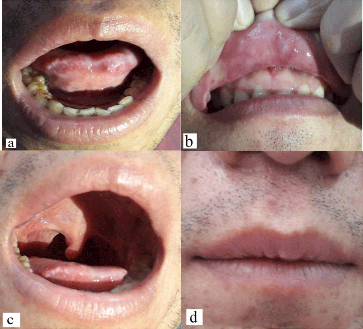
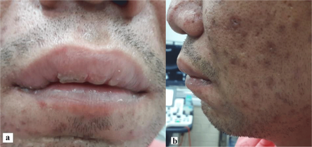
On physical examination, the patient was afebrile and vesicular crusting of chickenpox lesions had occurred. A diffuse swelling of the upper lip was noticed (Figure 1). It was soft in consistency, tender and non-pulsatile. Intraoral examination revealed multiple painful aphthous ulcers involving the tongue, labial mucosa and the anterior pillar of fauces with no regional lymphadenopathy (Figure 2). The remainder of clinical examination was unremarkable. Swabs were taken from the oral ulcers for the microbiological analysis.

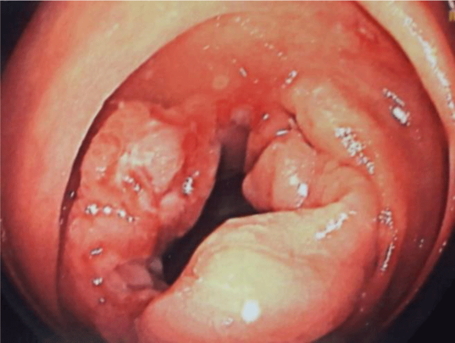
His complete blood count showed mild leukocytosis, with white blood cell count of 11.78 x 10⁹/L (normal range 4 - 11). Biochemical tests showed the following information: CRP 67.9 mg/L (normal range < 6), procalcitonin 0.03 ng/mL (normal range < 0.2), albumin 3.6 g/dL (normal range 3.5 – 5.5), iron 7 μmol/L (normal range 12.5 -25), transferrin saturation 16.6% (normal range 20 – 50), ferritin 332.4 ng/mL (normal range 20 – 400), folate 11.5 ng/mL (normal range 5.3 – 14), vitamin B12 902 pg/mL (normal range 211 - 911). Other biochemical profiles were within the reference range. Microbiological result was negative for HIV. Cytomegalovirus (CMV) and Epstein-Barr virus (EBV) polymerase chain reactions on blood samples were also negative. Oral swab culture was polymicrobial. The patient underwent colonoscopy which showed a large ulcer at the right colon (Figure 3). Multiple biopsies were performed. The histopathology conclusion was chronic colitis with ulceration.
During diagnostic workup, topical antiseptic therapy (Betadine mouth wash solution three times per day), local analgesics (i.e. lidocaine 2%), and systemic corticosteroid (methylprednisolone 40 mg per day) with proton pump inhibitor were prescribed. However, after 10 days of treatment, the patient did not show any improvement. The oral lesions were so painful that he could only take liquid food. Therefore, a biopsy specimen of oral ulcers was advised to identify the exact etiology. Under local anesthesia, a biopsy at the site of the anterior pillar of fauces was performed.
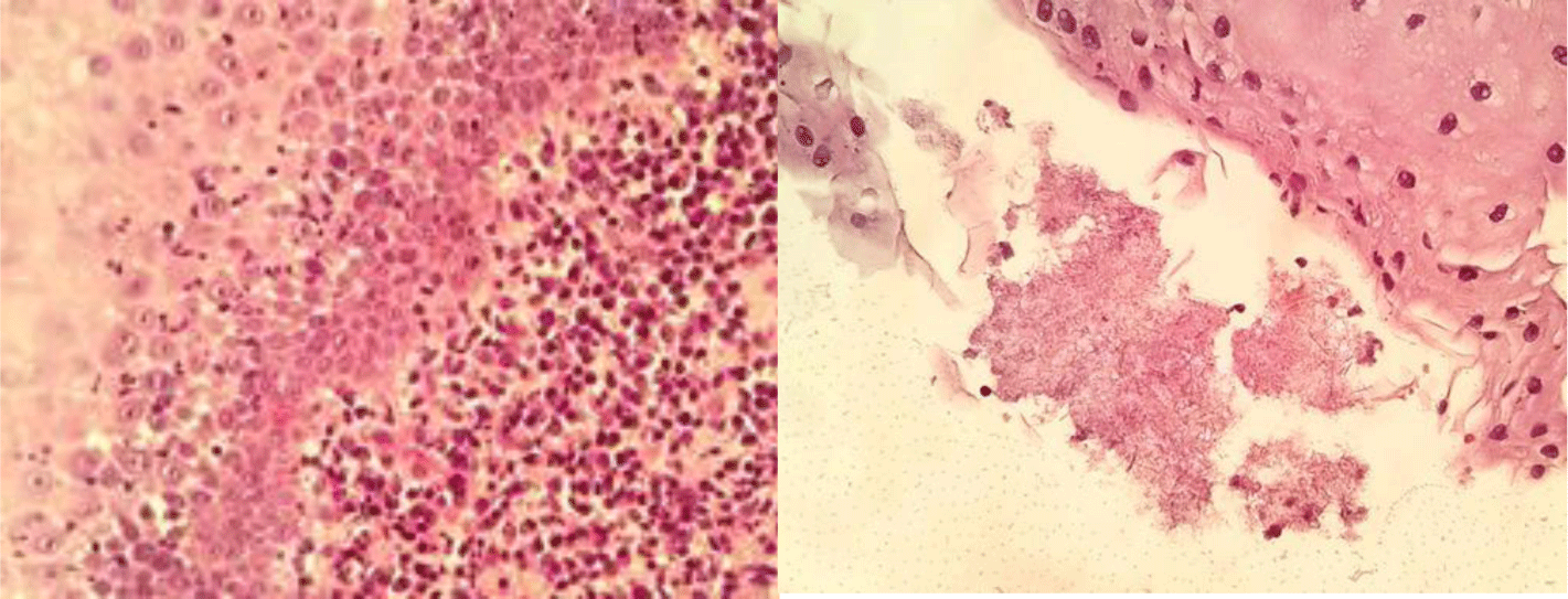
Thereafter, infliximab at a dose of 5 mg/kg was started to treat colonic Crohn’s disease and possible oral Crohn’s disease as adalimumab did not promote colonic mucosal healing in this patient. After a week, the patient denied any improvement on his oral lesions. The histopathology report came back showing chronic tonsillitis with Actinomyces spp. (Figure 4). A regimen of amoxicillin 1g three times a day was added and the patient experienced a marked response after 2 weeks (Figure 5).


3. DISCUSSION
Oral lesions in adult CD patients have a prevalence rate ranging between 20% and 50%. Aphthous ulcers, the most common type of oral lesions, occurring in 20–30% of adult patients with CD. Oral localisation in CD is characterised by a marked male predominance, a young age at onset and a very protracted course. Common symptoms include pain on touch or on eating acidic or spicy foods, impaired eating, speaking, swallowing and, of importance, psychological distress and even depression due to altered cosmetic appearance [4].
It is difficult to determine the exact cause of oral lesions i.e., oral manifestation of CD itself or related to CD therapy (drug reactions, infections) or other aetiologies. Therefore, a thorough physical examination and diagnostic work-up including microbiological and pathological profiles should be performed. In this case, histopathology report showed Actinomyces colonies on the surface of the tonsil.
The causative agent of oral actinomycosis is originated from flora of the oropharyngeal mucous membrane. Disruption of the mucosal barrier is the main triggering factor of the infection. Actinomycosis of the oral cavity can be present as a mass or abscess or ulcerative lesion or sinus [10]. Actinomyces are very difficult to grow in culture with <30% diagnostic yield, which limits the usefulness of microbiological identification in such infections [11]. Similarly, other laboratory findings like anemia, leukocytosis, and an increase in ESR are non-specific and are not supportive to establish the diagnosis.
Gram staining of pus and pathology of infected tissue are of great interest for the diagnosis of actinomycosis, as it is usually more sensitive than culture. Once Actinomyces spp. have invaded tissues, they develop a chronic granulomatous infection characterized by the formation of tiny clumps, called sulfur granules because of their yellow color. Histopathology examination discloses one to three sulfur granules in about 75% of cases, described as basophilic masses with eosinophilic terminal clubs on staining with hematoxylin and eosin (Splendore–Hoeppli phenomenon) [7]. The biopsy we obtained was small, therefore no Splendore–Hoeppli reaction was found.
We found only 5 published cases of actinomycosis with known CD including 3 cases of abdominal actinomycosis [12-14] and 2 cases of pulmonary actinomycosis [15, 16]. To our knowledge, no case of oral actinomycosis has previously been reported. In 3 out of 5 published cases, the patients were under biologics [14-16]. Our patient was under adalimumab before admission. Immunocompromised hosts are at increased risk for infection with Actinomyces [14, 16]. Actinomycosis was diagnosed by culture in 2 cases [12, 14], by histopathology in 2 cases [13, 16], and by both tests in 1 case [15]. Culture and pathology are keys to diagnose actinomycosis.
Actinomyces spp. are usually extremely susceptible to beta-lactams, especially penicillin G or amoxicillin. As a consequence, penicillin G or amoxicillin is considered drug of choice for the treatment of actinomycosis. Patients with actinomycosis require prolonged (6- to 12-month) high doses of penicillin G or amoxicillin to facilitate the drug penetration in abscess and in infected tissues [7]. Our patient showed a marked response after 2 weeks of high dose amoxicillin. Therefore, he would continue this regimen for up to 6 months.
Conclusion
In conclusion, this study reports a case of actinomycosis of the oral cavity in a patient with CD, effectively treated with oral amoxicillin. It is important to consider actinomycosis in the differential diagnosis of oral lesions in CD patients, particularly in immunosuppressed patients. Bacterial cultures and pathology are the cornerstones of diagnosis. Patients with actinomycosis require prolonged (6- to 12-month) high doses of penicillin G or amoxicillin, until the lesions are completely healed.


