Original article

Comparison of indocyanine green clearance test and Child-Pugh score in evaluation of pre-hepatectomy liver function

Viet Quoc Danga,b,*https://orcid.org/0000-0003-3545-9850, Quang Tien Phamahttps://orcid.org/0000-0001-8661-8554, Nghia Phuoc Phanbhttps://orcid.org/0000-0001-8313-0174, Phu Hong Phama,bhttps://orcid.org/0000-0002-6617-7381, Dat Tien Lea,bhttps://orcid.org/0000-0002-5623-9737, Thuan Duc Nguyenbhttps://orcid.org/0000-0003-3450-8673, Long Cong Duy Trana,bhttps://orcid.org/0000-0002-3994-4864, Thao Thi Phuong Doanc,dhttps://orcid.org/0000-0003-2181-3417, Bac Hoang Nguyena,bhttps://orcid.org/0000-0002-2973-7909
Author Information & Copyright
aDepartment of Surgery, Faculty of Medicine, University of Medicine and Pharmacy at Ho Chi Minh City, Ho Chi Minh city, Vietnam
bDepartment of Hepatobiliary and Pancreatic Surgery, University Medical Center Ho Chi Minh City, Ho Chi Minh city, Vietnam
cDepartment of Pathology, Faculty of Medicine, University of Medicine and Pharmacy at Ho Chi Minh City, Ho Chi Minh city, Vietnam
dDepartment of Pathology, University Medical Center Ho Chi Minh city, Ho Chi Minh city, Vietnam
*Address correspondence: Viet Quoc Dang at the Department of Surgery, Faculty of Medicine, University of Medicine and Pharmacy at Ho Chi Minh City, 217 Hong Bang street, Ward 11, District 5, Ho Chi Minh city, Vietnam; E-mail: viet.dq@umc.edu.vn

© Copyright 2022 MedPharmRes. This is an Open-Access article distributed under the terms of the Creative Commons Attribution Non-Commercial License (http://creativecommons.org/licenses/by-nc/4.0/) which permits unrestricted non-commercial use, distribution, and reproduction in any medium, provided the original work is properly cited.

Received: Dec 07, 2021; Revised: Jan 15, 2021; Accepted: Jan 25, 2022

Published Online: Oct 30, 2022

Abstract:

Introduction:

Indocyanine green (ICG) clearance (through ICG retention rate at 15 minutes - ICG-R15) is proven to correlate with histological fibrosis stage. Child-Pugh score, although proven to have weaker correlation, is still one of pre-hepatectomy liver function assessments. This study is to compare ICG-R15 and Child-Pugh score in evaluation of histological fibrosis stage and predicting of post-hepatectomy liver failure (PHLF) and to create the model of staging estimation for fibrosis.

Methods:

A prospective cohort study was conducted in 340 patients of hepatectomy. ICG-R15, Child-Pugh score and platelet count (PLT) were analyzed to examine their association with histological fibrosis stage and PHLF. Ordinal logistic regression was used to establish the model of staging estimation for fibrosis.

Results:

Child-Pugh score showed no significant association with histological fibrosis stage (p = 0.257) while ICG-R15 had a weak correlation (r = 0.232, p < 0.001), INR had a weak correlation (r = 0.156, p = 0.004), PLT had a negative correlation (r = -0.378, p < 0.001). The histological fibrosis stage could be estimated based on gender, age, ICG-R15 and PLT with AUC of 0.68. ICG-R15 was shown to be related to PHLF (p = 0.039) in which non-PHLF group had 0.75 times lower ICG-R15 than PHLF group while Child-Pugh score was shown to be statistically insignificant.

Conclusion:

ICG clearance test was better than Child-Pugh score in evaluation of pre-hepatectomy liver function and predicting of PHLF. It was possible to estimate the histological fibrosis stage based on gender, age, ICG-R15 and PLT.

Keywords: Indocyanine green clearance; ICG-R15; hepatectomy; post-hepatectomy liver failure; PHLF

1. INTRODUCTION

Evaluation of pre-hepatectomy liver function is a very important step to predict and prevent severe PHLF which has low incidence of 0 - 5.1% (1-7) but high mortality rate, up to 75% (2). Although Child-Pugh score was shown to have insignificant correlation to histological fibrosis stage in Child-Pugh A patients (8), this is still one of the criteria of assessing pre-hepatectomy liver function (9, 10).

ICG-R15 is considered as one of the best liver function tests and applied worldwide, especially in Asia - Pacific regions and becomes one of the standards for decision of the indication and the extent of liver resection (9, 11-13).

ICG-R15 has a close correlation to liver function deficiency and portal hypertension (14-18) as well as the progression to decompensated cirrhosis (19). Almost all patients undergoing hepatectomies are in Child-Pugh class A. For those patients, ICG-R15 is demonstrated to have a weak correlation to the histological staging of chronic hepatitis (Laennec’s staging system) (20) while Child-Pugh A class has an insignificant correlation to Ishak fibrosis scoring system (8).

ICG-R15 is shown to be associated with PHLF (21-24) and is used to guide the extent of hepatectomies (11) but some researches show that ICG-R15 is not an independent factor in predicting of PHLF (6).

ICG clearance test helps surgeons to predict the histological fibrosis stage in order to make a safe decision of hepatectomies, especially major hepatectomies (over 3 segmentectomies). Therefore, we carry out this study to compare ICG clearance test and Child-Pugh score in evaluating of histological fibrosis stage (Ishak fibrosis staging system) and predicting of PHLF as well as create the model of staging estimation for fibrosis from liver function tests.

2. MATERIALS AND METHOD

2.1. Patients

This was a prospective cohort study from October 2016 to March 2021 at University Medical Center - Ho Chi Minh city. This article was one of the results of the research “The role of Indocyanine green test in evaluation of pre-hepatectomy liver function” which was accepted and allowed by the Ethical Board of Biomedical research of University of Medicine and Pharmacy at Ho Chi Minh city on July 9th 2019, ID 316/ÐHYD-HÐÐÐ.

All patients with ICG clearance test results before hepatectomies for malignant or benign liver tumors or donor, were included in the study. We excluded those with bile obstruction or chemotherapy within 1 month because these conditions render ICG clearance test results inaccurate (25, 26). This study included 340 patients.

2.2. Data collection

All patients’ demographic data (gender, age), status of hepatitis, liver function tests (international normalized ratio of prothrombin time (INR), total serum bilirubin, serum albumin (Child-Pugh score)) and the platelet count (PLT) were collected. Pre-hepatectomy ICG clearance test was performed in all patients by LiMON method. After intravenous injection of indocyanine green 0.5 mg/kg, the indocyanine green disappearance rate was calculated by linear regression from the plasma concentrations of indocyanine green at 5, 10, and 15 minutes that gave us the two results: plasma disappearance rate (ICG-PDR) and ICG retention rate at 15 minutes (ICG-R15).

The operative factors (operation time, estimated blood loss, operative procedure) and the post-operative factors (histological staging of chronic hepatitis, tumor characteristics), liver function tests at post-operative day 3, 5, 7 (for PHLF diagnosis and grading), morbidity, mortality and hospital stay were recorded.

Histological staging of chronic hepatitis was based on Ishak fibrosis staging system for post-hepatectomy liver parenchyma (Ishak score) (27, 28).

PHLF was diagnosed and graded by International Study Group of Liver Surgery (ISGLS) criteria. PHLF is defined as a postoperatively acquired deterioration in the ability of the liver to maintain its synthetic, excretory, and detoxifying functions, which are characterized by an increased INR and concomitant hyperbilirubinemia on or after postoperative day 5 (29).

Child-Pugh score was calculated by 2 clinical features (ascites, hepatic encephalopathy) and 3 blood tests (serum albumin, serum bilirubin, INR). This score and ICG clearance were analyzed and compared in relation to histological fibrosis stage and PHLF.

2.3. Statistical analysis

Data was analyzed by IBM SPSS 26.0 and R 4.0.5. Continuous variables were described by quartiles and were compared by T-test and One-Way ANOVA (standard distribution) or Mann-Whitney U test and Kruskal-Wallis test (non-standard distribution). Nominal or ordinal variables were described by incidence and compared by Chi-Square test or Fisher’s Exact test.

Model of fibrosis staging was constructed based on ordinal logistic regression model, from univariate to multivariate model and reduced by backward stepwise variable selection based on AIC (Akaike Information Criterion). The index of effectiveness of the model was validated and optimism corrected by 1000-time bootstrap resampling.

3. RESULTS

3.1. Patient characteristics

There were 340 patients including 279 men (82.1%) and 61 women (17.9%). The patients’ characteristics were shown in Table 1 and Table 2.

Table 1. Pre-operative characteristics (n = 340)
Characteristics Median (Q1-Q3)/Number (%)
Age 59 (51 – 66)
BSA 1.66 (1.56 – 1.75)
Serum albumin (g/L) 40.60 (37.80 – 42.90)
Serum bilirubin (mmol/L) 12.20 (9.50 – 14.74)
INR 1.05 (1.00 – 1.11)
Platelet count (G/L) 214 (164 – 275)
ICG-PDR (%) 19.50 (16.60 – 22.70)
ICG-R15 (%) 5.40 (3.30 – 8.30)
Hepatitis
 Non-B non-C 82 (24.1)
 B 206 (60.6)
 C 51 (15.0)
 B and C 1 (0.3)
Child-Pugh score
 5 306 (90.0)
 6 31 (9.1)
 7 3 (0.9)
Number of TACE
 No 273 (80.3)
 1 time 50 (14.7)
 2 times 7 (2.1)
 ≥ 3 times 10 (2.9)
Liver hypertrophy
 None 300 (88.2)
 PVE (26 with previous TACE) 32 (9.4)
 1st phase ALPPS (1 with previous TACE) 8 (2.4)

BSA: body surface area. INR: international normalized ratio. ICG-PDR: ICG plasma disappearance rate. ICG-R15: ICG retention rate at 15 minutes. TACE: transcatheter arterial chemoembolization. PVE: portal vein embolization. ALPPS: associating liver partition and portal vein ligation for staged hepatectomy

Download Excel Table
Table 2. Operative and post-operative characteristics (n = 340)
Characteristics Median (Q1-Q3)/Number (%)
Operative time (minutes) 150 (120 – 180)
Blood loss (mL) 150 (100 – 200)
Hospital stay (days) 8 (7 – 10)
Hepatectomy
 Major (≥4 segments) 137 (40.3)
 Minor (< 4 segments) 203 (59.7)
Morbidity
 No 268 (78.8)
 PHLF 42 (12.4)
 Other 30 (8.8)
PHLF
 No 298 (87.6)
 Grade A 30 (8.8)
 Grade B 10 (2.9)
 Grade C 2 (0.6)
Death 3 (0.9)
Tumor nature
 HCC 270 (79.4)
 Necrotic tissue (after TACE) 2 (0.6)
 CCC 29 (8.5)
 HCC + CCC 1 (0.3)
 Liver metastasis 14 (4.0)
 Neuroendocrine tumor 1 (0.3)
 Benign tumor 14 (4.0)
 Normal liver parenchyma (liver donation) 9 (2.7)
Histological fibrosis stage (Ishak score)
 0/6 112 (32.9)
 1/6 53 (15.6)
 2/6 33 (9.7)
 3/6 54 (15.9)
 4/6 46 (13.5)
 5/6 36 (10.6)
 6/6 6 (1.8)

PHLF: post-hepatectomy liver failure. HCC: hepatocellular carcinoma. CCC: cholangiocellular carcinoma. TACE: transcatheter arterial chemoembolization. Other morbidity included biliary fistula, ascites, fluid collection, biliary stenosis, intraoperative biliary injury, pneumonia, acute renal injury, myocardial infraction

Download Excel Table
3.2. Comparison of ICG clearance test and Child-Pugh score in histological fibrosis stage evaluation

There was no significant difference of Ishak score between men and women (p = 0.148). Age had a weak correlation to Ishak score with Spearman’s rho of 0.167 significantly different from 0 (p = 0.002).

There were 80 patients with previous methods of treatment for liver hypertrophy before major hepatectomies including transcatheter arterial chemoembolization (TACE), portal vein embolization (PVE) (or both) or the 1st phase of associating liver partition and portal vein ligation for staged hepatectomy (ALPPS). These did not affect ICG-R15 (p = 0.141) and ICG-PDR (p = 0.138) as well as Ishak score (p = 0.074).

ICG-R15 had a weak correlation to Ishak score with Spearman’s rho of 0.232 significantly different to 0 (p < 0.001) (Table 3).

Table 3. Relationship between pre-operative characteristics and histological fibrosis stage (Ishak score)
Characteristics Spearman’s rho p
Gender 0.148
Age 0.167 0.002
Previous treatments 0.074
ICG-R15 0.232 < 0.001
Child-Pugh 0.257
Serum albumin -0.087 0.121
Serum bilirubin 0.104 0.061
INR 0.156 0.004
Platelet count -0.378 < 0.001

ICG-R15: ICG retention rate at 15 minutes. INR: international normalized ratio

Download Excel Table

There was no significant difference of Ishak score among 3 scores of Child-Pugh 5, 6, 7 (p = 0.257). Serum albumin and bilirubin had no correlation to Ishak score with Spearman’s rho of -0.087 (p = 0.121) and 0.104 (p = 0.061) respectively, while INR had a weak correlation to Ishak score with Spearman’s rho of 0.156 significantly different to 0 (p = 0.004).

Platelet count had a weak negative correlation to Ishak score with Spearman’s rho of -0.378, significantly different to 0 (p < 0.001).

3.3. Model of histological fibrosis stage estimation

Based on the demographical variables (gender, age) and pre-operative liver function tests (ICG-R15, serum bilirubin, INR, platelet count), three models for histological staging estimation for fibrosis were constructed using the ordinal logistic regression model:

  • - Model 1: univariate of ICG-R15

  • - Model 2: multivariate of 6 upper variables

  • - Model 3: multivariate of 6 upper variables but reduced by backward stepwise variable selection based on Akaike Information Criterion (AIC)

Table 4 showed estimated parameters from these models and effectiveness in Ishak score estimation. ICG-R15, INR and female had a positive correlation to Ishak score (OR > 1, p < 0.05) while PLT and serum bilirubin had a negative correlation to Ishak score (OR < 1, p > 0.05). PLT and serum bilirubin were not chosen in the reduced multivariate model. The index of effectiveness of the model was validated and optimism corrected by 1000-time bootstrap resampling.

Table 4. Model of histological fibrosis staging estimation
Univariate Multivariate Reduced multivariate
OR (95% CI) p OR (95% CI) p OR (95% CI) p
ICG-R15 (double increase) 3.73 (2.11-6.57) <0.001 1.36 (1.12-1.65) 0.001 2.75 (1.47-5.14) 0.001
Age (+1 year) - - 1.03 (1.01-1.04) 0.005 1.02 (1.00-1.04) 0.015
Sex: (Female to Male) - - 1.89 (1.00-2.93) 0.019 1.65 (0.97-2.81) 0.066
PLT (+10 G/L) - - 0.92 (0.89-0.94) <0.001 0.92 (0.89-0.94) <0.001
Serum bilirubin (+10 mmol/L)) - - 0.89 (0.59-1.34) 0.677 - -
INR (+0.1) - - 1.23 (0.97-1.56) 0.075 - -
Effectiveness (AUC) 0.59 0.68 0.68

OR: odd ratio for higher Ishak score when variables increase one unit or compare to standard value. CI: confidence interval. ICG-R15: ICG retention rate at 15 minutes. PLT: platelet count. INR: International Normolized Ratio. AUC: area under the curve

Download Excel Table

The effectiveness of each model shows that the multivariate analysis produced more accurate estimation than the univariate one. In spite of having less than 2 variables, the reduced multivariate model still produced the equivalent effectiveness to the full multivariate one. Between two patients with different Ishak score, the reduced multivariate model could identify the patient with the higher score with 69% accuracy.

In clinical practice, the estimating equation for Ishak score from the parameters in Table 5 or the simplified nomogram from the model could be used (Figure 1).

Table 5. Parameters from the reduced model
Parameters Values
α1 (to estimate P(Ishak ≥1)) 0.73241
α2 (to estimate P(Ishak ≥2)) -0.05656
α3 (to estimate P(Ishak ≥3)) -0.50158
α4 (to estimate P(Ishak ≥4)) -1.34980
α5 (to estimate P(Ishak ≥5)) -2.35889
α6 (to estimate P(Ishak ≥6)) -4.44481
β1 1.01106
β2 0.02192
β3 0.50060
β4 -0.08498
Download Excel Table
mpr-6-3-22-g1
Figure 1. Nomogram for estimating of Ishak score
Download Original Figure

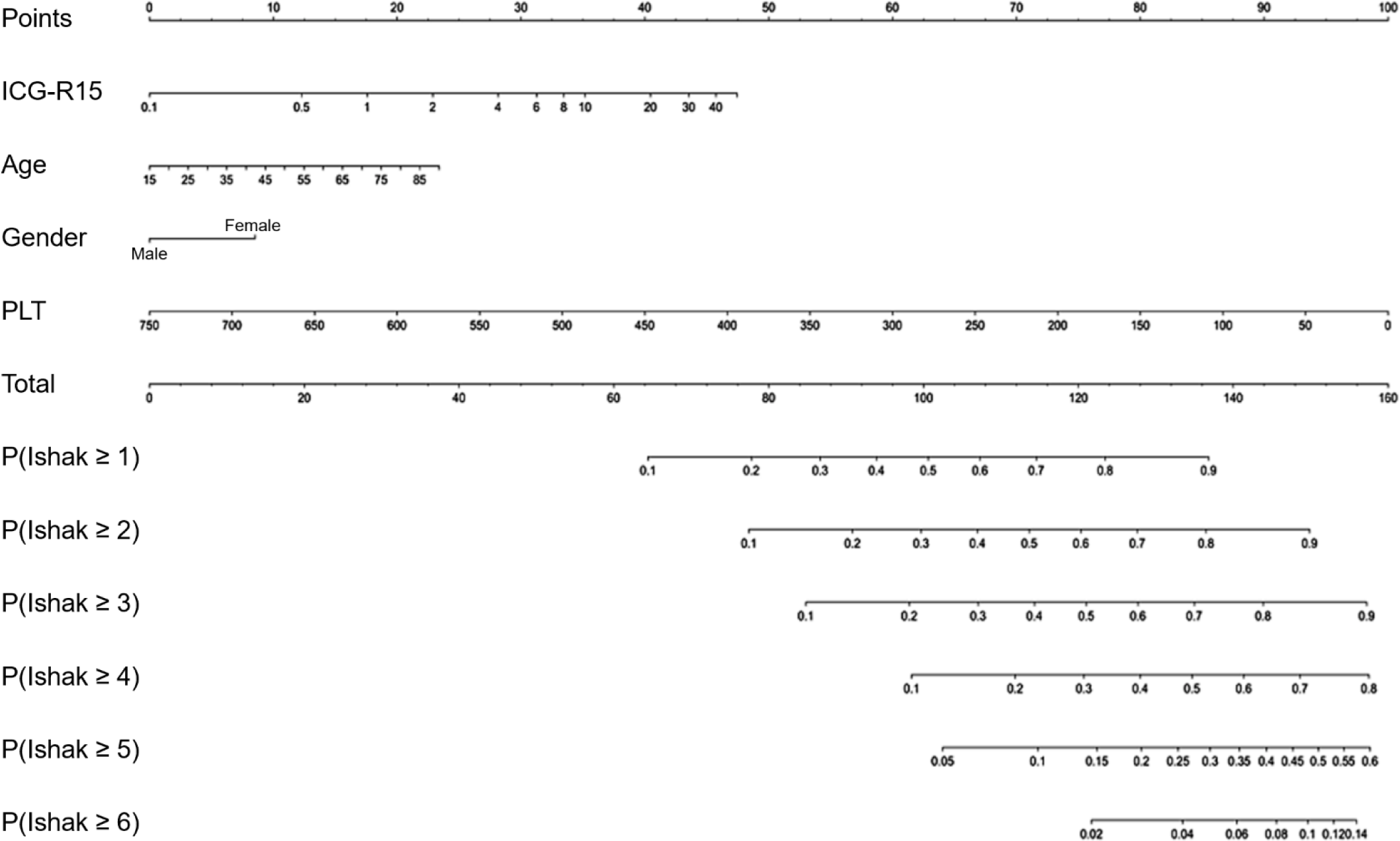
These steps could be followed to calculate the probability of Ishak score: (1) use the parameters in Table 5 and the value of ICG-R15, age, gender (Female = 1, Male = 0), PLT to calculate the 6 probabilities of Ishak score ≥ n from following equation, (2) combine the 6 probabilities and clinical experience to decide the histological fibrosis stage of the patients.

P ( Ishak n ) = plogis ( α n + β 1 × log 2 ( ICG R 15 ) + β 2 × Age + β 3 × Gender + β 4 × ( PLT / 10 ) )

The process of calculation could be simplified as follows: (1) calculate the points of every variable based on the values of ICG-R15, age, gender, PLT, then calculate the total points, (2) calculate the 6 probabilities P (Ishak ≥ n) and combine them with clinical experience to decide the histological fibrosis stage of the patients.

3.4. Comparison of ICG clearance test and Child-Pugh score in predicting of PHLF

ICG clearance significantly was associated with PHLF (p = 0.019). ICG-R15 value in non-PHLF group is 0.75 times lower than PHLF-group (95% CI: 0.56 - 0.99). However, ICG-R15 was not associated with PHLF grading (Table 6).

Table 6. Relationship between liver function tests and PHLF
Non-PHLF (n= 298) PHLF (n = 42) p
ICG-R15 (%) 5.20 (3.10 – 8.23) 6.20 (4.25 – 9.53) 0.039
Serum albumin (g/L) 40.60 (37.95 – 42.98) 39.10 (36.40 – 41.58) 0.108
Serum bilirubin (mmol/L) 12.20 (9.50 – 14.67) 12.62 (9.45 – 18.98) 0.136
INR 1.05 (1.00 – 1.12) 1.06 (0.99 – 1.11) 0.864
Platelet count (G/L) 214 (165 – 278) 216 (129 – 266) 0.296

Statistics are median (Q1-Q3); PHLF: post-hepatectomy liver failure. ICG-R15: ICG retention rate at 15 minutes. INR: international normalized ratio

Download Excel Table

Child-Pugh score was not associated with PHLF (p = 1.000). Component tests of Child-Pugh score (including serum albumin, serum bilirubin, INR) and platelet count were not associated with PHLF significantly (p = 0.108, 0.136, 0.864, 0.296 respectively).

4. DISCUSSION

ICG-R15 is the most useful test for Asia - Pacific surgeons to evaluate pre-hepatectomy liver function and guide the majority of hepatectomies (9, 11) as well as predict PHLF (6, 21-24, 30). In this study, the median value of ICG-R15 was 5.4% (3.3 - 8.3%) which was good and common for hepatectomy.

The analysis of 80 patients with preceding treatments before hepatectomy, such as TACE or PVE or 1st phase ALPPS, showed that these treatments did not affect ICG-R15 and Ishak score and therefore, these did not affect analysis of the correlation of these two variables.

ICG-R15 had a weak correlation with Ishak score (r = 0.232) which corresponded another research using Laennec’s histological staging of chronic hepatitis system among Child-Pugh A patients with r = 0.325 (20). Although the correlation coefficient was low, surgeons still had a useful tool to estimate the histological fibrosis stage before hepatectomy. Child-Pugh score did not significantly relate to the Ishak fibrosis stage which concurred our hospital’s previous study (8). This might result from the fact that most of the patients in this study had Child-Pugh class A. This proved that good Child-Pugh score did not mean good liver parenchyma, which made it difficult for surgeons to make decisions of hepatectomy. Therefore, ICG clearance test was more valuable than Child-Pugh score in evaluation of histological fibrosis stage among patients indicated for hepatectomies. However, ICG-R15 was weaker than platelet count and stronger than INR in the correlation with Ishak score. This suggests that there was no best single test to predict the histological fibrosis stage, hence, the model of histological staging estimation for fibrosis was constructed.

The model was set up based on variables from the general knowledge and clinical experience of the relationship between cirrhosis and ICG-R15, age, gender, serum bilirubin, INR, platelet count. Ishak score was the ordinal variable (based on the histological staging); therefore, we could not estimate the score directly based on calculating but just on the probabilities of every stage. Surgeons should combine the probabilities of stages and clinical experience to decide which stage is suitable for the patient. Although the accuracy of this model was quite low, at 68%, it was already useful for surgeons to determine the indication and the extent of liver resection because it enabled the prediction of histological fibrosis stage for approximately 2/3 patients of hepatectomies.

As regards post-operative results, ICG-R15 in non-PHLF group was 0.75 times lower than PHLF group. In the absolute value of ICG-R15, this was not the large gap, but the difference of 25% was significantly notable. However, ICG-R15 did not significantly relate to PHLF grading. It is presumably because the quantities of PHLF grade B-C is low in this study. So, this study could not demonstrate the role of ICG-R15 in prognosticating the severity of PHLF. There was no evidence that Child-Pugh score and component tests as well as platelet count relate to the PHLF and its grades. In summary, ICG-R15 was better than Child-Pugh score in predicting of PHLF. This finding was similar to the result of Wang et al (24) which demonstrated that ICG-R15 was better than Child-Pugh score in prediction of PHLF.

This study had a quite large sample of 340 patients with all stages of histological fibrosis and cirrhosis and all kinds of hepatectomy which made the results more reliable and able to apply to clinical practice. Besides, this is one of very few studies about the relationship between liver function tests and histological fibrosis stage. However, comparing the continuous variable (ICG-R15) and the ordinal variable (Child-Pugh score) in correlation with another ordinal variable (Ishak score) was very challenging. Based on the clinical experience, we separated the component tests Child-Pugh scoring system and added platelet count to analyze the correlation to Ishak score to clarify the roles of the traditional liver function tests with the new one. In spite of this limitation, we still hoped this study would help surgeons have more information about pre-hepatectomy liver function assessments.

Conclusion

ICG clearance test, through ICG-R15 had a weak correlation to Ishak fibrosing score with r = 0.232. Child-Pugh score did not significantly correlate to Ishak fibrosing score while platelet count had a weak negative correlation with r = -0.378. It was possible to estimate the Ishak fibrosing score based on gender, age, ICG-R15 and platelet count with the accuracy 68%. ICG clearance test was also better than Child-Pugh (and component tests) in predicting of PHLF.

FUNDING

The authors received no financial support for the research, authorship, and/or publication of this article.

CONFLICT OF INTEREST

The authors declare that there is no conflict of interest.

REFERENCES

1.

Egeli T, Unek T, Agalar C, Ozbilgin M, Derici S, Cevlik AD, et al. The Analysis of Posthepatectomy Liver Failure Incidence and Risk Factors Among Right Liver Living Donors According to International Study Group of Liver Surgery Definition. Transplant Proc. 2019; 51(4):1121-6

2.

Navarro JG, Yang SJ, Kang I, Choi GH, Han DH, Kim KS, et al. What are the most important predictive factors for clinically relevant posthepatectomy liver failure after right hepatectomy for hepatocellular carcinoma?. Ann Surg Treat Res. 2020; 98(2):62-71

3.

Nguyễn Thị Mỹ Xuân An, Trần Công Duy Long. Application of liver volume measurement using computed tomography to hepatectomy. Journal of Medicine of Ho Chi Minh. 2012; 16(1):22-5.

4.

Phan Phước Nghĩa, Trần Công Duy Long, Phạm Hồng Phú. Outcomes of right hepatectomy for treatment of hepatocellular carcinoma. Journal of Medicine of Ho Chi Minh. 2019; 23(1):184-8.

5.

Trần Công Duy Long, Nguyễn Hoàng Bắc, Nguyễn Ðức Thuận, Lê Tiần Ðạt, Ðặng Quốc Việt, Phạm Hồng Phú. et al. Laparoscopic liver resection for hepatocellular carcinoma. Journal of Medicine of Ho Chi Minh. 2015; 19(1):225-33.

6.

Yamamoto G, Taura K, Ikai I, Fujikawa T, Nishitai R, Kaihara S, et al. ALPlat criterion for the resection of hepatocellular carcinoma based on a predictive model of posthepatectomy liver failure. Surgery. 2020; 167(2):410-6

7.

Ye JZ, Mai RY, Guo WX, Wang YY, Ma L, Xiang BD, et al. Nomogram for prediction of the international study Group of Liver Surgery (ISGLS) grade B/C Posthepatectomy liver failure in HBV-related hepatocellular carcinoma patients: an external validation and prospective application study. BMC Cancer. 2020; 20(1):1036

8.

Lê Tiến Ðạt, Nguyễn Ðức Thuận, Ðặng Tâm, Trần Công Duy Long. The correlation between Child-Pugh A and staging leasions in hepatocellular carcinoma. Journal of Medicine of Ho Chi Minh. 2011; 15(1):67-70.

9.

Bryant R, Laurent A, Tayar C, van Nhieu JT, Luciani A, Cherqui D. Liver resection for hepatocellular carcinoma. Surg Oncol Clin N Am. 2008; 17(3):607-33 ix. Epub 2008/05/20. doi:

10.

Truty MJ, Vauthey J-N. Surgical resection of high-risk hepatocellular carcinoma: patient selection, preoperative considerations, and operative technique. Annals of surgical oncology. 2010; 17(5):1219-25

11.

Imamura H, Sano K, Sugawara Y, Kokudo N, Makuuchi M. Assessment of hepatic reserve for indication of hepatic resection: decision tree incorporating indocyanine green test. J Hepatobiliary Pancreat Surg. 2005; 12(1):16-22

12.

Kobayashi Y, Kiya Y, Sugawara T, Nishioka Y, Hashimoto M, Shindoh J. Expanded Makuuchi’s criteria using estimated indocyanine green clearance rate of future liver remnant as a safety limit for maximum extent of liver resection. HPB (Oxford). 2019; 21(8):990-7

13.

Lee S-G, Hwang S. How I do it: assessment of hepatic functional reserve for indication of hepatic resection. 2005; 12(1):38-43

14.

Asenbaum U, Kaczirek K, Ba-Ssalamah A, Ringl H, Schwarz C, Waneck F, et al. Post-hepatectomy liver failure after major hepatic surgery: not only size matters. Eur Radiol. 2018; 28(11):4748-56

15.

Lisotti A, Azzaroli F, Buonfiglioli F, Montagnani M, Cecinato P, Turco L, et al. Indocyanine green retention test as a noninvasive marker of portal hypertension and esophageal varices in compensated liver cirrhosis. Hepatology. 2014; 59(2):643-50

16.

Lu HS, Hsin IF, Chen PH, Yang TC, Chang CY, Huang YH, et al. The indocyanine green retention test as a noninvasive marker for esophageal varices in patients with hepatocellular carcinoma. J Chin Med Assoc. 2020; 83(8):737-42

17.

Moller S, la Cour Sibbesen E, Madsen JL, Bendtsen F. Indocyanine green retention test in cirrhosis and portal hypertension: Accuracy and relation to severity of disease. J Gastroenterol Hepatol. 2018

18.

Pind ML, Bendtsen F, Kallemose T, Moller S. Indocyanine green retention test (ICG-r15) as a noninvasive predictor of portal hypertension in patients with different severity of cirrhosis. Eur J Gastroenterol Hepatol. 2016; 28(8):948-54

19.

Lisotti A, Azzaroli F, Cucchetti A, Buonfiglioli F, Cecinato P, Calvanese C, et al. Relationship between indocyanine green retention test, decompensation and survival in patients with Child-Pugh A cirrhosis and portal hypertension. Liver Int. 2016; 36(9):1313-21

20.

Gu J, Zhang E, Liang B, Zhang Z, Chen X, Huang Z. Effectiveness comparison of indocyanine green retention test with the cirrhotic severity scoring in evaluating the pathological severity of liver cirrhosis in patients with hepatocellular carcinoma and Child-Pugh grade A liver function. World J Surg Oncol. 2020; 18(1):79

21.

Au K-P, Chan S-C, Chok KS-H, Chan AC-Y, Cheung T-T, Ng KK-C, et al. Child-Pugh Parameters and Platelet Count as an Alternative to ICG Test for Assessing Liver Function for Major Hepatectomy. HPB Surg. 2017; 2017:2948030

22.

de Liguori Carino N, O’Reilly DA, Dajani K, Ghaneh P, Poston GJ, Wu AV. Perioperative use of the LiMON method of indocyanine green elimination measurement for the prediction and early detection of post-hepatectomy liver failure. Eur J Surg Oncol. 2009; 35(9):957-62

23.

Tomimaru Y, Eguchi H, Gotoh K, Kawamoto K, Wada H, Asaoka T, et al. Platelet count is more useful for predicting posthepatectomy liver failure at surgery for hepatocellular carcinoma than indocyanine green clearance test. J Surg Oncol. 2016; 113(5):565-9

24.

Wang YY, Zhao XH, Ma L, Ye JZ, Wu FX, Tang J, et al. Comparison of the ability of Child-Pugh score, MELD score, and ICG-R15 to assess preoperative hepatic functional reserve in patients with hepatocellular carcinoma. J Surg Oncol. 2018; 118(3):440-5

25.

Stockmann M, Malinowski M, Lock JF, Seehofer D, Neuhaus P. Factors influencing the indocyanine green (ICG) test: additional impact of acute cholestasis. Hepatogastroenterology. 2009; 56(91-92):734-8

26.

Wakiya T, Kudo D, Toyoki Y, Ishido K, Kimura N, Narumi S, et al. Evaluation of the Usefulness of the Indocyanine Green Clearance Test for Chemotherapy-Associated Liver Injury in Patients with Colorectal Cancer Liver Metastasis. Annals of Surgical Oncology. 2014; 21(1):167-72

27.

Ishak K, Baptista A, Bianchi L, Callea F, De Groote J, Gudat F, et al. Histological grading and staging of chronic hepatitis. Journal of Hepatology. 1995; 22(6):696-9

28.

Knodell RG, Ishak KG, Black WC, Chen TS, Craig R, Kaplowitz N, et al. Formulation and application of a numerical scoring system for assessing histological activity in asymptomatic chronic active hepatitis. Hepatology. 1981; 1(5):431-5

29.

Rahbari NN, Garden OJ, Padbury R, Brooke-Smith M, Crawford M, Adam R, et al. Posthepatectomy liver failure: A definition and grading by the International Study Group of Liver Surgery (ISGLS). Surgery. 2011; 149(5):713-24

30.

Honmyo N, Kobayashi T, Kuroda S, Oshita A, Onoe T, Kohashi T, et al. A novel model for predicting posthepatectomy liver failure based on liver function and degree of liver resection in patients with hepatocellular carcinoma. HPB : the official journal of the International Hepato Pancreato Biliary Association. 2021; 23(1):134-43