Original article

Naso-pharyngeal carriage and antimicrobial susceptibility of Streptococcus pneumoniae in community-acquired pneumonia in children

Phong Thi Nam Nguyenahttps://orcid.org/0000-0002-5197-8266, Julien M. Buyckbhttps://orcid.org/0000-0003-0299-4654, Stephen Bakerc,dhttps://orcid.org/0000-0003-1308-5755, Trang Hoang Thu Nguyenehttps://orcid.org/0000-0002-0818-3281, Tin Viet Phamahttps://orcid.org/0000-0001-5676-0226, Tuoi Thi Hong Dof,*https://orcid.org/0000-0002-5445-0694
Author Information & Copyright
aFaculty of Pharmacy, Danang University of Medical Technology and Pharmacy, Da Nang, Vietnam
bINSERM U1070, UFR de Médecine Pharmacie, Université de Poitiers, Poitiers, France
cCambridge Institute of Therapeutic Immunology and Infectious Disease, University of Cambridge School of Clinical Medicine, Cambridge Biomedical Campus, Cambridge, UK
dDepartment of Medicine, University of Cambridge School of Clinical Medicine, Cambridge Biomedical Campus, Cambridge, UK
eOxford University Clinical Research Unit, Hospital for Tropical Diseases, Ho Chi Minh City, Vietnam
fFaculty of Pharmacy, University of Medicine and Pharmacy at Ho Chi Minh City, Ho Chi Minh City, Vietnam
*Address correspondence: Tuoi Thi Hong Do at the Faculty of Pharmacy, University of Medicine and Pharmacy at Ho Chi Minh City, Ho Chi Minh City, Vietnam; E-mail: hongtuoi@ump.edu.vn

© Copyright 2022 MedPharmRes. This is an Open-Access article distributed under the terms of the Creative Commons Attribution Non-Commercial License (http://creativecommons.org/licenses/by-nc/4.0/) which permits unrestricted non-commercial use, distribution, and reproduction in any medium, provided the original work is properly cited.

Received: Jul 01, 2021; Revised: Feb 11, 2022; Accepted: Feb 15, 2022

Published Online: Dec 31, 2022

Abstract:

Introduction:

Streptococcus pneumoniae is the most common cause of community-acquired pneumonia (CAP) in children. Recently, the rapid emergence of antibiotic-resistant pneumococci has posed enormous challenges for CAP treatment and public health. This study aims to provide clinicians with updated data about the antimicrobial susceptibility of S. pneumoniae and improve the treatment guidelines in CAP in children.

Methods:

This cross-sectional and in-vitro study was conducted at three hospitals in Quang Nam province and Da Nang city in Vietnam. Pneumococcal strains were isolated from nasopharyngeal aspirate samples of 360 CAP in-patients under five years of age. The susceptibility of clinically used antibiotics was investigated using the disk diffusion test and the E-test for identifying the MIC. Multi-drug resistant (MDR) S. pneumoniae isolates were also determined.

Results:

21.9% of CAP children patients were pneumococcal carriage. The susceptibility testing demonstrated that all 56 tested pneumococcal isolates were resistant to erythromycin and azithromycin, whereas none of the isolates developed the resistance to levofloxacin, vancomycin, and linezolid. The clindamycin- and cotrimoxazole-resistant pneumococci rates were 96.4% and 87.5%, respectively. For β-lactam antibiotics, the resistance proportions of pneumococcal isolates to penicillin G, co-amoxiclav, amoxicillin, cefotaxime, and ceftriaxone were 3.6%,1.8%, 1.8%, 1.8%, and 3.6%, respectively. 83.9% of the tested isolates were MDR S. pneumoniae.

Conclusion:

Our data support using penicillin G, amoxicillin, co-amoxiclav, or cefotaxime as the first-line therapy for uncomplicated-pneumococci-induced CAP in Quang Nam – Da Nang, while vancomycin, linezolid, and levofloxacin should be used as alternatives or in MDR cases.

Keywords: CAP; Streptococcus pneumoniae; antimicrobial susceptibility; resistance; multi-drug resistance

1. INTRODUCTION

Community-acquired pneumonia (CAP) is the leading cause of death among many infectious diseases, with more than 1.6 million deaths worldwide annually, of which 0.7-1 million cases of children under five years of age [1, 2]. From a report to the United Nations Children’s Fund (UNICEF) and World Health Organization (WHO) in 2004, the Ministry of Health of Vietnam estimated about 4500 children under five years of age died due to CAP annually [3]. The most common causative agent of CAP was Streptococcus pneumoniae, which is usually found in the nasopharyngeal carriages and causes the highest mortality rate among pathogens [4-6]. Empirical therapy for nonsevere pneumonia in children was recommended for an antimicrobial regimen including amoxicillin or ampicillin to provide good coverage of S. pneumoniae [7].

In recent years, epidemiological surveys showed that antibiotic resistance had been a global problem, becoming one of the urgent health challenges for the next decades [8]. The resistance rate of S. pneumoniae to macrolides and many other antibiotics has considerably increased, especially in Asian countries [9, 10]. In Vietnam, the proportion of erythromycin-resistant S. pneumoniae was 92.2%, the highest among 11 Asian countries [9]. In 2011, also in Vietnam, pneumococcal isolates from respiratory infections developed resistance to broad-spectrum antibiotics such as cotrimoxazole (91%), tetracycline (78.6%), chloramphenicol (67.9%), and second-generation cephalosporins such as cefuroxime (71.4%), cefaclor (87.6%) [11].

Due to the overuse of antibiotics and the rapid increase in antimicrobial resistance, it is necessary to revise the guidelines regularly to gain clinical effectiveness and lower the clinical failures, mortality rate, and the burden of treatment costs on the patients [12, 13]. In addition, because the resistance rate could vary according to healthcare settings, it should be investigated and continuously updated.

In line with the national action plan on antimicrobial resistance, we investigated the antimicrobial susceptibility of S. pneumoniae isolates collected from CAP children at three hospitals in Quang Nam province and Da Nang city from September 2017 to September 2018. This study aims to provide clinicians with critical and updated data on antibiotic resistance patterns and support them to select appropriately and safely antibiotics for empirical treatment of CAP.

2. MATERIALS AND METHODS

2.1. Study design

This cross-sectional and in-vitro study was conducted in the middle region of Vietnam at the pediatric departments of three hospitals, including Da Nang Hospital for Women and Children (DN) - a secondary referral hospital in central Vietnam; Quang Nam Hospital for Women and Children (QN1) - the representative hospital in the urban area of Quang Nam province; and General Hospital at Northern Mountain of Quang Nam (QN2) - the representative hospital in the rural area of Quang Nam province from September 2017 to September 2018. The number of beds of DN, QN1, and QN2 is about 500, 370, and 350, respectively. In QN1 and QN2, the beds can be added depending on the number of patients admitted to the hospital. Annually, about 4000 children are admitted to Da Nang Hospital for Women and Children due to acute respiratory infections, whereas this figure for Quang Nam Hospital for Women and Children and General Hospital at Northern Mountain of Quang Nam is approximately 3500.

2.2. Materials, samples, and study process
Sample size calculation for estimating the population proportion [14]
n = ( Z 1 α / 2 ) 2 x P ( 1 P ) / d 2 = 323

Where:

  • - n: Minimum sample size

  • - Z 1-α/2 : The number of standard errors away from the mean (Z 1-α/2 = 1.96 for a level of confidence of 95% or a level of significance of 5%)

  • - P: The estimated proportion of the population p = 0.3, based on previous research [15-17] (The pneumococcal nasopharyngeal carriage rate in children with CAP was 21.5%, 46%, 28.6%, respectively)

  • - d: The distance, in either direction, from the population proportion (d = 0.05)

The minimum sample size was calculated at 323 children and rounded to 360. The number of patents of each hospital was 120. The association between qualitative variables was analyzed with Pearson’s chi-square test using SPSS statistics software 20.0.

Sampling, isolation, identification of S. pneumoniae

Clinical specimens were collected by a convenient sampling method with the support of hospitals. Nasopharyngeal aspirates (NPA) of CAP inpatients from 2 months to 60 months who had not received antibiotics for more than 24 hours before hospitalization were obtained by nurses of the hospitals. The collection was performed based on the guidelines of the Vietnam Ministry of Health [18] and the Center for Disease Control and Prevention [19].

The samples were stored in sterile vials and labeled with the collection date, name, age, gender, and ID number of the patients, then transported to the hospital’s laboratory for culture and isolation.

The specimen was cultured on a Blood Agar (BA) plate with 5% sheep blood and then incubated at 35-37°C in 5% CO2 for 16-24 hours. The identification of pneumococcal isolates underwent steps such as observation of colony morphology, gram staining, optochin susceptibility testing, bile salt solubility [20]. The suspected strains were confirmed by Buker’s MALDI-TOF (matrix-assisted laser desorption/ionization - time-of-flight) mass spectrometry. S. pneumoniae strains were kept in Brain Heart Infusion (BHI) broth with a supplement of 5% sheep blood (manufactured by Nam Khoa. LTD) and 20% glycerin and stored at -70°C.

Determination of the antimicrobial susceptibility

The antimicrobial susceptibility testing (AST) of S. pneumoniae isolates were performed with 12 antimicrobials clinically used in the three hospitals by the disk diffusion method following guidelines established by the Clinical and Laboratory Standards Institute (CLSI) and, when required, by minimum inhibitory concentrations (MICs) by E-test [21]. Kirby-Bauer disk diffusion method was used for testing of erythromycin (E:15 μg disc), azithromycin (AZM:15 μg disc), clindamycin (DA: 2.0 μg disc), levofloxacin (LEV: 5.0 μg disc), trimethoprim + sulfamethoxazole (SXT: 25 μg disc), vancomycin (VA: 30 μg disc), linezolid (LZD: 30 μg disc) (Oxoid). For the other antimicrobials such as penicillin G (PG), amoxicillin (AC), amoxicillin + acid clavulanic (XL), cefotaxime (CT), ceftriaxone (TX), the MICs were identified by using E-test strips (BioMérieux).

The inoculum was prepared by direct colony suspension method using colonies from overnight-cultured BA plates. The turbidity of suspension was adjusted to 0.5 McFarland, and suspension should be used within 15 minutes after preparation. The suspension was streaked evenly over the surface of Mueller-Hinton blood agar (MHBA) plates using a sterile cotton swab. The plate cover was slightly opened for 5 minutes to absorb excess liquid into the agar fully. The antibiotic disc or E-Test strips was applied to the surface of the MHBA within 15 minutes of inoculation and then gently pressed to ensure that it was entirely in contact with the agar. The MHBA plates were incubated at 35-37°C in 5% CO2 for 18-24h [22-24]. Quality control of antibiotic discs and E test strips was performed using strain S. pneumoniae ATCC 49619 [21, 25].

Reading and interpretation of the results

After 18-24 hours of incubation, results were read by the reflected light method. The inhibition zones of the antibiotic discs were measured using a ruler or calipers. The MIC was identified at the point where the ellipse intersects the scale, and the growth lawn was distinct. If the intersection point fell between two-fold dilutions, the MIC value was rounded up to the highest value. If the intersection differs on either side of the strip, MIC was identified as the greater value [23]. MIC breakpoints or inhibition zones for defining interpretive susceptibility were published by the Clinical and Laboratory Standards Institute (CLSI) [21].

Multi-drug resistance (MDR) was defined as acquired resistance to at least one agent in three or more antimicrobial groups [26-29].

2.3. Statistical method

Data were processed by using excel version 16.49 and analyzed by using SPSS statistics software 20.0. Variables specific to the research population used such as hospitals (DN, QN1 and QN2); age groups (2-23 months and 24-60 months); gender (Male, female); culture (positive, negative); isolation (positive, negative). The association between qualitative variables was analyzed using Pearson’s chi-square test with a level of confidence of 95%.

2.4. Ethical consideration

The study was approved by the Danang Hospital for Women and Children, two Quang Nam Provincial Hospitals, and the Department of Health of Quang Nam Province (approval decision No 3287/QÐ-UBND on September 08, 2017). Accordingly, the hospitals supported our research on clinical specimens and the isolation and identification of pneumococcus.

3. RESULTS

3.1. Naso-pharyngeal carriage of S. pneumoniae in children with CAP

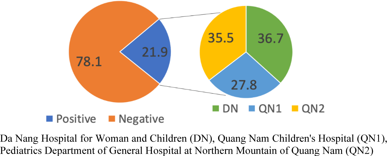
Overall, among 360 children with CAP in our study, there was a significantly higher prevalence of CAP in males than females (61.1% vs. 38.9%, p < 0.001). The majority of CAP children (62.2%, 224/360) aged between 2 – 23 months, while 37.8% (136/360) of cases aged from 24 to 60 months with significant difference (p < 0.001). In general, the prevalence of pneumococcal pneumonia (culture-confirmed with S. pneumoniae) was 21.9% (79/360 CAP cases). There was no significant difference among the three hospitals with the respective prevalence of pneumococcal pneumonia in each hospital were 24.2%, 18.3%, and 23.3% in DN, QN1, and QN2 (p = 0.498) (Table 1). As of 79 S. pneumoniae isolates, strains collected at QN2 and DN took up higher proportions (36.7% and 35.55, respectively) of the total pneumococcal isolates, while QN1 occupied a lower proportion, 27.8% (Figure 1). There was no significant difference in the S. pneumoniae-infected rate between the three hospitals (p = 0.498), between 2 age groups (p = 0.571), and also between two genders (p = 0.217).

Table 1. Prevalence of pneumococcus in isolates and cultures
Culture Isolation
Positive N (%) Negative N (%) p-value Positive N (%) Negative N (%) p-value
Hospital
  DN 29/120 (24.2) 91/120 (75.8) 0.498 29/45 (64.4) 16/45 (35.6) 0.366
  QN1 22/120 (18.3) 98/120 (81.7) 22/43 (51.2) 21/43 (48.8)
  QN2 28/120 (23.3) 92/120 (76.7) 28/44 (63.5) 16/44 (36.4)
Age group in months
  2-23 47/224 (21.0) 177/224 (79.0) 0.571 47/77 (61.0) 30/77 (39.0) 0.741
  (24-60) 32/136 (23.5) 104/136 (76.5) 32/55 (58.2) 23/55 (41.8)
Gender
  Males 53/220 (24.1) 167//220 (75.9) 0.217 53/88 (60.2) 35/88 (39.8) 0.900
  Females 26/140 (18.6) 114/140 (81.4) 26/44 (59.1) 18/44 (40.9)

Da Nang Hospital for Woman and Children (DN), Quang Nam Children’s Hospital (QN1), Pediatrics Department of General Hospital at Northern Mountain of Quang Nam (QN2)

Download Excel Table
mpr-6-4-34-g1
Figure 1. Positive rate for S. pneumoniae in 3 hospitals
Download Original Figure
3.2. The susceptibility of S. pneumoniae isolates on clinically used antimicrobials

A total number of 79 S. pneumoniae strains was initially isolated from 360 CAP cases. However, only 56 S. pneumoniae strains were tested for antimicrobial susceptibility. The missing was due to various reasons, including cell death (caused by freezer malfunction, autolysis during the transportation from Da Nang to OUCRU lab in Ho Chi Minh), strains lost, or contamination.

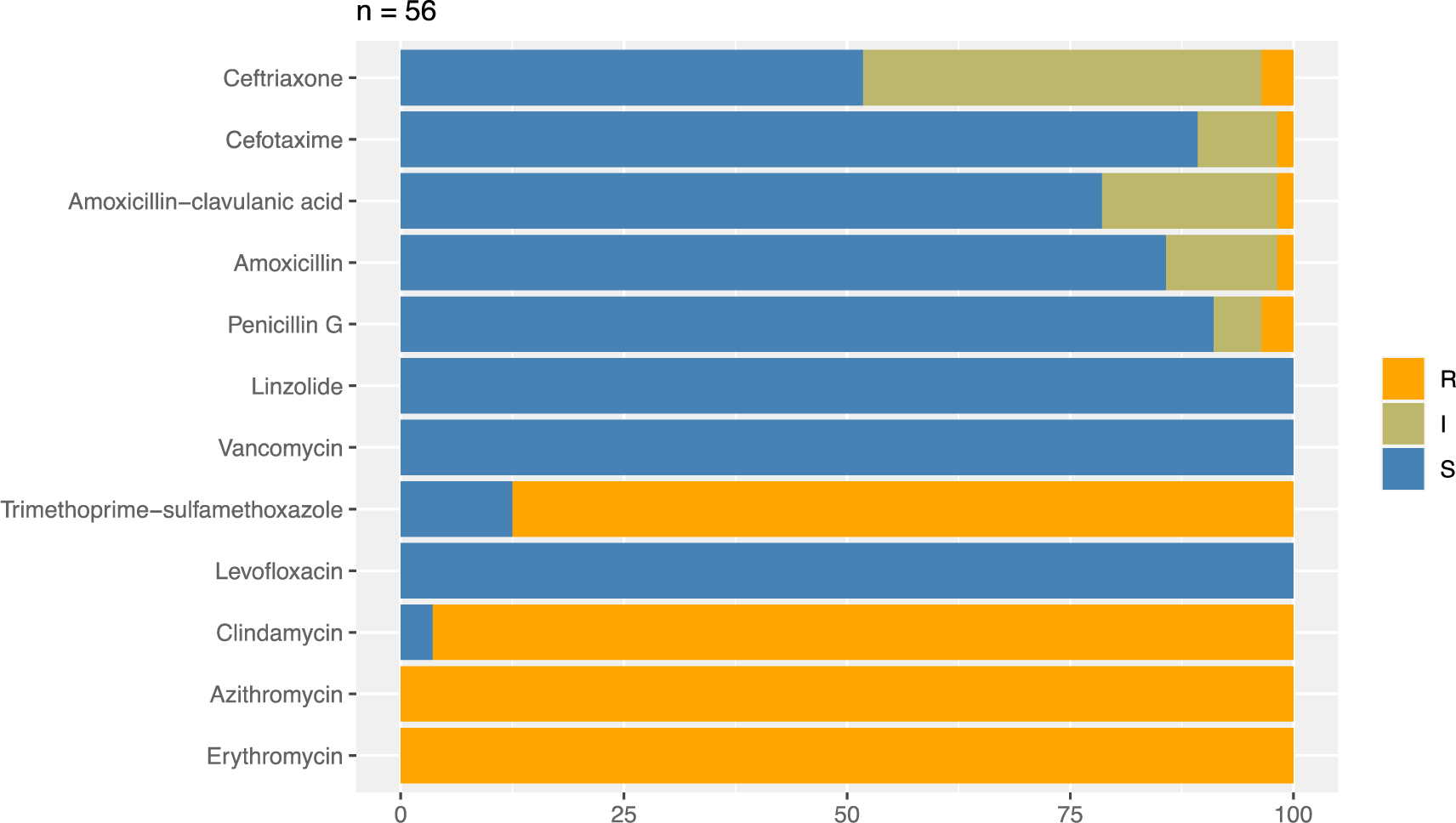
Regarding the AST results, a hundred percent of S. pneumoniae isolates (56 isolates) were resistant to macrolides, including erythromycin and azithromycin. Also, the high prevalence of pneumococci isolates was resistant to clindamycin and sulfamethoxazole-trimethoprim at 96.4 and 87.5%, respectively. Conversely, CAP S. pneumoniae isolates showed a very low resistance rate to β-lactams including penicillin G (3.6%), amoxicillin-clavulanate (1.8%), amoxicillin (1.8%), cefotaxime (1.8%), and ceftriaxone (3.6%). Especially, all S. pneumoniae in the study were susceptible to levofloxacin, vancomycin, and linezolid (Figure 2).

mpr-6-4-34-g2
Figure 2. Percentage of susceptible-mediate-resistant S. pneumoniae strains to antibiotics. (S: Susceptible; I: Intermediate; R: Resistant, n: number of S. pneumoniae strains)
Download Original Figure
The minimum inhibitory concentration of five antimicrobials to S. pneumoniae

MICs of penicillin G, amoxicillin-clavulanate, amoxicillin, cefotaxime, and ceftriaxone for 56 tested pneumococcal isolates were identified by the gradient method using E- test and demonstrated in table 2.

Table 2. MIC distribution of 56 pneumococcal strains for antibiotics
The number of strains with MIC values corresponding to the antibiotic and cumulative percent
MIC value μg/mL Penicillin G Amoxicillin Amoxicillin/ Clavulanic acid Cefotaxime Ceftriaxone
n (c)% n (c)% n (c)% n (c)% n (c)%
0.008-0.094 2 3.6 3 5.4 3 5.4 3 5.4 1 1.8
0.125-0.5 14 28.6 12 26.8 13 28.6 32 62.5 13 25.0
0.75-0.94 6 39.3 9 42.9 5 37.5 8 76.8 7 37.5
1 9 55.4 9 58.9 7 50.0 7 89.3 8 51.8
1.5 13 78.6 10 76.8 11 69.6 3 94.6 12 73.2
2 7 91.1 5 85.7 5 78.6 2 98.2 13 96.4
3 2 94.6 4 92.9 8 92.9 0 98.2 0 96.4
4 1 96.4 3 98.2 2 96.4 0 98.2 0 96.4
6 0 96.4 0 98.2 1 98.2 0 98.2 2 100
8 1 98.2 0 98.2 0 98.2 0 98.2 0 100
12-16 1 100 1 100 1 100 1 100 0 100
24-256 0 100 0 100 0 100 0 100 0 100
Strains in total 56 56 56 56 56

MIC: Minimum Inhibitory Concentration, (n): number of strains, (c) %: cumulative percent Green: Susceptible; Yellow: Intermediate; Orange: Resistant

Download Excel Table

The MICs of penicillin G and amoxicillin for S. pneumoniae isolates ranged from 0.016 to 12 μg/mL, in which 91.1% of S. pneumoniae isolates with penicillin MIC ≤ 2.0 μg/mL and 92.9% with amoxicillin MIC ≤ 3.0 μg/mL.

For amoxicillin + acid clavulanic (co-amoxiclav), the range of MICs on 56 S. pneumoniae isolates was between 0.016 and 16 μg/mL. The MICs below three μg/mL were observed in 93% of the total isolates.

For the remaining two antimicrobials, the result found that 89.3% of pneumococcal isolates exhibited cefotaxime MIC ≤ 1.0 μg/mL while the rate of strains with ceftriaxone MIC ≤ 2.0 μg/mL was 96.4%.

The proportion of MDR S. pneumoniae isolates at three hospitals

Overall, the proportion of MDR S. pneumoniae was 83.9% (47/56 isolates). In particular, the proportion of MDR S. pneumoniae at each hospital, including DN, QN1, and QN2, was 77.3% (17/22 isolates), 86.7% (13/15 isolates), and 89.5% (17/19 isolates), respectively. There was no significant difference between these three hospitals in the rate of CAP caused by MDR S. pneumoniae (p = 0.538).

4. DISCUSSION

Naso-pharyngeal carriage of S. pneumoniae in children with community-acquired pneumonia

In diagnoses of lower respiratory tract infections, NPA specimens were commonly used in many studies with high specificity [30-33] and less invasive in children [34]. Regarding the recovery rate, NPA indicated the highest rate of isolates of S. pneumoniae (33%) while oral pharyngeal swab (OPS) had the lowest rate with 20% [30]. The rate of detection of S. pneumoniae in NPA specimens collected from patients with CAP in our study was 21.9%. This proportion was similar to the result of the survey of Negash (21.5%) [15] and much higher than that of a study carried out on children aged from 0 to 12 months with CAP in China (13.9%) [33] and South Korea (2%) [32].

The discrepancy between pneumococcal carriage rates of patients may be influenced by the patient’s exposure to antibiotics before sample collection. It is difficult to determine the exact recall history and the detection of antibiotics in urine samples [15]. Children who had been treated with antibiotics before swabbing had significantly lower rates of S. pneumoniae than patients who had not received prior antibiotics [35]. Besides, techniques used to detect the presence of pneumococci in specimens also affected the results. For example, culture methods might show lower positive cases than PCR (polymerase chain reaction) [36].

In-vitro antimicrobial activity on S. pneumoniae isolated from clinical specimens

Our results in figure 2 have demonstrated that all isolates of S. pneumoniae are entirely resistant to macrolides, including erythromycin and azithromycin. The trend of macrolide resistance is increasing compared to previous years in Vietnam. In 2004 and 2012, the rate of drug resistance of S. pneumoniae to erythromycin was 92.1% and over 99%, respectively [9, 11]. The prevalence of macrolide-resistant pneumococcal isolates in Vietnam is relatively similar to other Asian countries [31-33]. Such a high resistance rate could be attributed to the majority of resistance genes among pneumococcal strains in Asian countries [37], especially the erm(B) gene in 97.9% of macrolide-resistant strains of S. pneumoniae [10]. This gene encodes ribosomal methylase that can dimethylate the subunit of 23S rRNA, which is the main target site of macrolides [38]. This resistance mechanism also enables S. pneumoniae to develop resistance to other antibiotics, such as lincosamides. This may be an explanation for the high rate of isolates resistant to clindamycin in our study (96.4%) and the previous survey in Vietnam (85%) [11].

In the face of the high level of antimicrobial resistance, whether macrolides or clindamycin are still clinically effective for treating CAP caused by S. pneumoniae remains a controversial question. There may be some differences between in vitro and in vivo results due to the influence of host-related factors such as the immune system, the nature and severity of the infection, and the type and dose of the infection. Numerous reports state that macrolides enhance the efficacy of combination therapy, especially with β-lactam antibiotics [39-41]. Some authors suggest that the clinical benefit of macrolides does not come from their antibacterial properties but rather their anti-inflammatory effects. The anti-inflammatory effects of macrolides are illuminated by their ability to inhibit the production of interleukin-8 of bronchial epithelial cells [42]. In addition, macrolides can inhibit protein synthesis and reduce virulence factors [43, 44]. Therefore, adding macrolides may provide a better antibacterial spectrum, especially atypical agents.

On the other hand, our study found that 100% of S. pneumoniae isolates were susceptible to vancomycin, linezolid, and levofloxacin. This result is accordant with surveys carried out previously in Vietnam and Asian countries [11, 45, 46]. Therefore, using vancomycin in treating respiratory infections caused by multi-drug resistant pneumococcal isolates would undoubtedly produce good outcomes. However, it should be noticed that there was the emergence of vancomycin-tolerant S. pneumoniae isolates in some countries [47, 48]. Therefore, vancomycin should be prescribed as an alternative therapy in the cases caused by penicillin-resistant pneumococcal isolates (MIC ≥ 4.0 μg/mL) [49].

Regarding linezolid and levofloxacin, a study on pneumococcal isolates collected worldwide illustrated that 100% of S. pneumoniae strains were susceptible to these antibiotics [50]. In the guidelines of IDSA (Infectious Diseases Society of America), linezolid and levofloxacin are considered to use as alternatives or in the case of penicillin-resistant pneumococcal isolates (MIC ≥ 4.0 μg/mL) [49]. However, linezolid and levofloxacin could trigger severe adverse drug reactions (ADRs). Hence, they are absent in some guidelines for children, such as the British National Formulary (BNF) [51], European guidelines [52], and Canadian Paediatric Society [7].

Our study revealed that most S. pneumoniae strains isolated from 3 hospitals were still susceptible to β-lactam antibiotics. In Vietnam, the rate of β-lactam resistant S. pneumoniae has still been relatively lower than in some Asian countries, such as China and Korea [53, 54]. The resistance mechanism of S. pneumoniae to β-lactams is the modification of PBPs (Penicillin-binding proteins), decreasing the affinity of β-lactams with their targets rather than the production of beta-lactamase [55].

It is worthy of note that there is no difference between the resistance rate to amoxicillin and co-amoxiclav. Therefore, in CAP’s case caused by S. pneumoniae, amoxicillin should be prescribed instead of co-amoxiclav to lower the treatment cost.

Noticeably, the data showed that cefotaxime demonstrated a better antimicrobial effect than ceftriaxone in the cephalosporin group. The susceptibility rate of pneumococcal isolates to cefotaxime was 89.3%, whereas this percentage for ceftriaxone was 51.8%. The ceftriaxone MIC of 96.4% strains was ≤ 2 μg/mL. The increase of ceftriaxone non-susceptible S. pneumoniae isolates in the present could be explained by the emergence and combination of mutations on three resistance genes, PBP1a, 2b, and 2x, especially PBP 2b, which is responsible for encoding ceftriaxone low-affinity PBPs [56, 57].

Nowadays, multidrug-resistant S. pneumoniae is an issue of concern as well. Our research also showed that 47 of 56 pneumococcal strains were resistant to multiple antibiotics, representing 83.9%. In Vietnam, among 84 strains isolated from patients under five years old with lower respiratory tract infections, the rate of S. pneumoniae resistance to one or more antibiotics was 96%, 78% of which was multidrug-resistant pneumococci [58]. Based on a study in Thailand in 2014, the multidrug-resistance rate of S. pneumonia was 31.6% [59]. Pneumococcal MDR has many causes, primarily due to widespread use and abuse of antibiotics in the community, unnecessary prescribing by physicians and patients who do not adhere to the regime [60].

Until now, β-lactams have shown a powerful antimicrobial effect on S. pneumoniae isolates in Da Nang city and Quang Nam province. Hence, they should be the first choice for treating CAP in children. However, in cases of reduced susceptibility to β-lactams antibiotics, dose adjustment is required based on pharmacokinetics/pharmacodynamics, and antimicrobial therapy must be used reasonably to not increase the future resistance rate.

In addition to the appropriate use of antibiotics in clinical settings, the widespread use of pneumococcal conjugate vaccines such as PCV-7 or PCV-13 is significantly essential to lower the antimicrobial resistance of S. pneumoniae. The introduction of PCV7 and PCV-13 in the USA in the first decade of the 21st has remarkably reduced antibiotic-resistant pneumococcal infections [61]. Although the emergence of non-vaccine serotypes might reduce the effectiveness of pneumococcal vaccines [61, 62], they have still played a pivotal role in fighting pneumococcal diseases and tackling the problem of antibiotic resistance. Therefore, PCV-7 and PCV-13 should be introduced in the Expanded Vaccination Program in Vietnam.

Our study has some limitations. The only nasopharyngeal aspirate specimen was used to isolate the organisms. As the nature of a fastidious organism (S. pneumoniae) that was vulnerable to changing temperature, most of the missing strains in our study were due to cell death during the transportation in long distances.

Conclusion

Our study showed updated data of CAP children caused by S. pneumoniae in Da Nang and Quang Nam. It would be significantly beneficial for further studies, particularly the investigations on the effectiveness of PCV, to design and run an appropriate vaccination program.

Our data advocate the use of penicillin G, amoxicillin, co-amoxiclav, or cefotaxime as the first-line therapy for uncomplicated S. pneumonia-induced CAP in children Quang Nam – Da Nang. Vancomycin, linezolid, and levofloxacin should be used as alternatives or for the infections caused by multi-drug resistant S. pneumoniae. Due to a high prevalence of macrolide-resistant S. pneumoniae, a macrolide empirical monotherapy is not recommended for the management of CAP unless atypical microorganisms are suspected. Because the prevalence of multidrug-resistant pneumococci in Da Nang and Quang Nam (83.9%) is an issue of concern, it is required to make a dramatic change in the antibiotic-prescribing habits of physicians, other health care workers, and the public about the proper and wise use of antibiotics.

FUNDING

The authors received no financial support for the research, authorship, and/or publication of this article.

CONFLICT OF INTEREST

The authors declare that there is no conflict of interest.

ACKNOWLEDGEMENTS

The authors would like to thank Professor William Couet - Director of the unit of Pharmacology of Antimicrobial agents, Dr. Julien M. Buyck - INSERM U1070 for research orientation and conceptualization. We thank Professor Stephen Baker for supporting materials and facilities to conduct the study at Oxford University Clinical Research Unit, the Hospital for Tropical Diseases, Ho Chi Minh City, Vietnam. Besides, we thank Danang Hospital for Women and Children and two hospitals in Quang Nam for bacterial samples used in this study and Danang University of Medical Technology and Pharmacy to access their research facilities. All other contributions to this study should be acknowledged.

AUTHORS’ CONTRIBUTION

HTTD, JMB developed the concept and critical review the manuscript. NPTN was responsible for study design and sample collection, conducted the experiment, and drafted the manuscript with TTHN and TVP ‘s support. All members contributed to data analysis, interpretation, critical review and approved the final manuscript.

REFERENCES

1.

Ferreira-Coimbra J, Sarda C, Rello J. Burden of Community-Acquired Pneumonia and Unmet Clinical Needs. Advances in therapy. 2020; 37(4):1302-18

2.

World Health Organization Department of Maternal Newborn Child and Adolescent Health ib, World Health Organization pb. Revised WHO classification and treatment of pneumonia in children at health facilities : evidence summaries. Switzerland: Geneva: World Health Organization. 2014.

3.

Ministry of Health of Vietnam. Decision on Issuing Guidelines for community pneumonia management in children. No101 / QD-BYT. Ha Noi 2014; .

4.

Gerard Tortora BF. Christine Case. Microbiology: an introduction. 13th ed United States of America: Pearson. 2018 January; 8.

5.

Mathur S, Fuchs A, Bielicki J, Van Den Anker J, Sharland M. Antibiotic use for community-acquired pneumonia in neonates and children: WHO evidence review. Paediatrics and international child health. 2018; 38(sup1):S66-s75

6.

Cillóniz C, Ewig S, Polverino E, Marcos MA, Esquinas C, Gabarrús A, et al. Microbial aetiology of community-acquired pneumonia and its relation to severity. Thorax. 2011; 66(4):340-6

7.

Le Saux N, Robinson JL. Uncomplicated pneumonia in healthy Canadian children and youth: Practice points for management. Paediatrics & child health. 2015; 20(8):441-50

8.

WHO. Antimicrobial resistance. 2020 [[updated 13 October 2020]]. Available from: https://www.who.int/news-room/fact-sheets/detail/antimicrobial-resistance.

9.

Song JH, Jung SI, Ko KS, Kim NY, Son JS, Chang HH, et al. High prevalence of antimicrobial resistance among clinical Streptococcus pneumoniae isolates in Asia (an ANSORP study). Antimicrobial agents and chemotherapy. 2004; 48(6):2101-7

10.

Kim SH, Song JH, Chung DR, Thamlikitkul V, Yang Y, Wang H, et al. Changing trends in antimicrobial resistance and serotypes of Streptococcus pneumoniae isolates in Asian countries: an Asian Network for Surveillance of Resistant Pathogens (ANSORP) study. Antimicrobial Agents and Chemotherapy. 2012; 56(3):1418-26

11.

Phạm HV, Đoàn MP, Trần TTT, Nam L, Nguyễn TNA, Nguyễn SMT, et al. Resistance to antibiotics of S. pneumoniae and H. influenzae isolated from acute respiratory infections - Results of multi-center research conducted in Vietnam (SOAR) 2010 - 2011. Journal of Practical Medicine. 2012; 885(12):6-11.

12.

Song JH, Thamlikitkul V, Hsueh PR. Clinical and economic burden of community-acquired pneumonia amongst adults in the Asia-Pacific region. International Journal of Antimicrobial Agents. 2011; 38(2):108-17

13.

Klugman KP. Bacteriological evidence of antibiotic failure in pneumococcal lower respiratory tract infections. The European Respiratory Journal Supplement. 2002; 36:3s-8s

14.

Lemeshow S, Hosmer DW, Klar J, Lwanga SK. WHO. Adequacy of sample size in health studies. Chichester : Wiley: World Health Organization. 1990.

15.

Negash AA, Asrat D, Abebe W, Hailemariam T, Gebre M, Verhaegen J, et al. Pneumococcal Carriage, Serotype Distribution, and Risk Factors in Children With Community-Acquired Pneumonia, 5 Years After Introduction of the 10-Valent Pneumococcal Conjugate Vaccine in Ethiopia. Open forum infectious diseases. 2019; 6(6):ofz259

16.

Lahti E, Peltola V, Waris M, Virkki R, Rantakokko-Jalava K, Jalava J, et al. Induced sputum in the diagnosis of childhood community-acquired pneumonia. Thorax. 2009; 64(3):252-7

17.

Singh M, Agarwal A, Das RR, Jaiswal N, Ray P. Naso-pharyngeal Carriage of Organisms in Children Aged 3-59 months Diagnosed with Severe Community-acquired Pneumonia. Indian pediatrics. 2016; 53(2):125-8

18.

Trần ĐP. Instructions for sampling, packaging, preservation and transportation of infectious disease specimens. Ha Noi: Ministry of Health - Preventional Medical Department. 2017.

19.

CDC. Specimen collection guidelines. 2020.Available from: https://www.cdc.gov/urdo/downloads/SpecCollectionGuidelines.pdf.

20.

World Health Organization & Centers for Disease Control and Prevention (U.S.). Laboratory methods for the diagnosis of meningitis caused by neisseria meningitidis, Streptococcus pneumoniae, and Haemophilus influenzae. WHO manual. 2nd edWorld Health Organization. 2011; 74p.

21.

CLSI. Performance Standards for Antimicrobial Susceptibility Testing; Twenty-fifth Informational Supplement. CLSI document M100-S25. Wayne, PA: Clinical and Laboratory Standards Institute. 2015.

22.

Ministry of Heath of Vietnam. Practical Guidelines of Clinical Microbiology Testing Techniques (Issued together with Decision No. 1539 / QD-BYT dated April 20, 2017 of the Minister of Health). 2017; 197:243.

23.

BioMérieux. ETest- Antimicrobial Susceptibility Testing. 2012.Available from: https://www.ilexmedical.com/files/E-test-Package-Insert/AST_WW.pdf.

24.

Hudzicki J. Kirby-Bauer Disk Diffusion Susceptibility Test Protocol: American Society for Microbiology. 2009 [[cited 2018 Wed, 29 Aug]]. Available from: https://asm.org/getattachment/2594ce26-bd44-47f6-8287-0657aa9185ad/Kirby-Bauer-Disk-Diffusion-Susceptibility-Test-Protocol-pdf.pdf.

25.

Biodisk AB. Etest application sheet, S. pneumoniae. 2007.Available from: https://www.ilexmedical.com/files/Etest-ApplicationSheets/streppneumoniae.pdf.

26.

Kim L, Mc Gee L, Tomczyk S, Beall B. Biological and Epidemiological Features of Antibiotic-Resistant Streptococcus pneumoniae in Pre- and Post-Conjugate Vaccine Eras: a United States Perspective. Clinical microbiology reviews. 2016; 29(3):525-52

27.

Golden AR, Rosenthal M, Fultz B, Nichol KA, Adam HJ, Gilmour MW, et al. Characterization of MDR and XDR Streptococcus pneumoniae in Canada, 2007-13. The Journal of Antimicrobial Chemotherapy. 2015; 70(8):2199-202

28.

Sallam M, Abbadi J, Natsheh A, Ababneh NA, Mahafzah A, Özkaya Şahin G. Trends in Antimicrobial Drug Resistance of Streptococcus pneumoniae Isolates at Jordan University Hospital (2000-2018). Antibiotics (Basel, Switzerland). 2019; 8(2)

29.

Cillóniz C, Garcia-Vidal C, Ceccato A, Torres A. Antimicrobial Resistance Among Streptococcus pneumoniae.In: In: Fong IW, Shlaes D, Drlica K, editors.editors Antimicrobial Resistance in the 21st Century. Cham: Springer International Publishing. 2018; p p. 13-38

30.

Rapola S, Salo E, Kiiski P, Leinonen M, Takala AK. Comparison of four different sampling methods for detecting pharyngeal carriage of Streptococcus pneumoniae and Haemophilus influenzae in children. Journal of Clinical Microbiology. 1997; 35(5):1077-9

31.

Capeding MR, Nohynek H, Sombrero LT, Pascual LG, Sunico ES, Esparar GA, et al. Evaluation of sampling sites for detection of upper respiratory tract carriage of Streptococcus pneumoniae and Haemophilus influenzae among healthy Filipino infants. Journal of Clinical Microbiology. 1995; 33(11):3077-9

32.

Lee S, Lee K, Kang Y, Bae S. Prevalence of serotype and multidrug-resistance of Streptococcus pneumoniae respiratory tract isolates in 265 adults and 36 children in Korea, 2002-2005. Microbial Drug Resistance (Larchmont, NY). 2010; 16(2):135-42

33.

Yu YY, Xie XH, Ren L, Deng Y, Gao Y, Zhang Y, et al. Epidemiological characteristics of nasopharyngeal Streptococcus pneumoniae strains among children with pneumonia in Chongqing, China. Scientific reports. 2019; 9(1):3324

34.

Lu AZ, Shi P, Wang LB, Qian LL, Zhang XB. Diagnostic Value of Nasopharyngeal Aspirates in Children with Lower Respiratory Tract Infections. Chinese Medical Journal. 2017; 130(6):647-51

35.

Greenberg D, Broides A, Blancovich I, Peled N, Givon-Lavi N, Dagan R. Relative importance of nasopharyngeal versus oropharyngeal sampling for isolation of Streptococcus pneumoniae and Haemophilus influenzae from healthy and sick individuals varies with age. Journal of Clinical Microbiology. 2004; 42(10):4604-9

36.

Athlin S, Strålin K. The Binax NOW Streptococcus pneumoniae test applied on nasopharyngeal aspirates to support pneumococcal aetiology in community-acquired pneumonia. Scandinavian Journal of Infectious Diseases. 2013; 45(6):425-31

37.

Schroeder MR, Chancey ST, Thomas S, Kuo WH, Satola SW, Farley MM, et al. A Population-Based Assessment of the Impact of 7- and 13-Valent Pneumococcal Conjugate Vaccines on Macrolide-Resistant Invasive Pneumococcal Disease: Emergence and Decline of Streptococcus pneumoniae Serotype 19A (CC320) With Dual Macrolide Resistance Mechanisms. Clinical infectious diseases : an official publication of the Infectious Diseases Society of America. 2017; 65(6):990-8

38.

Schroeder MR, Stephens DS. Macrolide Resistance in Streptococcus pneumoniae. Frontiers in Cellular and Infection Microbiology. 2016; 6(98)

39.

Tessmer A, Welte T, Martus P, Schnoor M, Marre R, Suttorp N. Impact of intravenous {beta}-lactam/macrolide versus {beta}-lactam monotherapy on mortality in hospitalized patients with community-acquired pneumonia. The Journal of Antimicrobial Chemotherapy. 2009; 63(5):1025-33

40.

Martínez JA, Horcajada JP, Almela M, Marco F, Soriano A, García E, et al. Addition of a macrolide to a beta-lactam-based empirical antibiotic regimen is associated with lower in-hospital mortality for patients with bacteremic pneumococcal pneumonia. Clinical infectious diseases : an official publication of the Infectious Diseases Society of America. 2003; 36(4):389-95

41.

Metersky ML, Ma A, Houck PM, Bratzler DW. Antibiotics for bacteremic pneumonia: Improved outcomes with macrolides but not fluoroquinolones. Chest. 2007; 131(2):466-73

42.

Takizawa H, Desaki M, Ohtoshi T, Kawasaki S, Kohyama T, Sato M, et al. Erythromycin modulates IL-8 expression in normal and inflamed human bronchial epithelial cells. American Journal of Respiratory And Critical Care Medicine. 1997; 156(1):266-71

43.

Kaul R, McGeer A, Norrby-Teglund A, Kotb M, Schwartz B, O’ Rourke K, et al. Intravenous immunoglobulin therapy for streptococcal toxic shock syndrome--a comparative observational study. The Canadian Streptococcal Study Group. Clinical infectious diseases : an official publication of the Infectious Diseases Society of America. 1999; 28(4):800-7

44.

Caballero J, Rello J. Combination antibiotic therapy for community-acquired pneumonia. Annals of Intensive Care. 2011; 1:48

45.

Song JH. Emergence and spread of antimicrobial resistance of Streptococcus pneumoniae in Korea. Yonsei Medical Journal. 1998; 39(6):546-53

46.

Zhao C, Li Z, Zhang F, Zhang X, Ji P, Zeng J, et al. Serotype distribution and antibiotic resistance of Streptococcus pneumoniae isolates from 17 Chinese cities from 2011 to 2016. BMC Infectious Diseases. 2017; 17(1):804

47.

Novak R, Henriques B, Charpentier E, Normark S, Tuomanen E. Emergence of vancomycin tolerance in Streptococcus pneumoniae. Nature. 1999; 399(6736):590-3

48.

Moscoso M, Domenech M, García E. Vancomycin tolerance in clinical and laboratory Streptococcus pneumoniae isolates depends on reduced enzyme activity of the major LytA autolysin or cooperation between CiaH histidine kinase and capsular polysaccharide. Molecular Microbiology. 2010; 77(4):1052-64

49.

Bradley JS, Byington CL, Shah SS, Alverson B, Carter ER, Harrison C, et al. The Management of Community-Acquired Pneumonia in Infants and Children Older Than 3 Months of Age: Clinical Practice Guidelines by the Pediatric Infectious Diseases Society and the Infectious Diseases Society of America. Clinical Infectious Diseases. 2011; 53(7):e25-e76

50.

Zhang Z, Chen M, Yu Y, Pan S, Liu Y. Antimicrobial susceptibility among Streptococcus pneumoniae and Haemophilus influenzae collected globally between 2015 and 2017 as part of the Tigecycline Evaluation and Surveillance Trial (TEST). Infection and Drug Resistance. 2019; 12:1209-20

51.

PaediatricFormularyCommittee. BNF for children. London: Pharmaceutical Press. 20192020.

52.

Esposito S, Cohen R, Domingo JD, Pecurariu OF, Greenberg D, Heininger U, et al. Antibiotic therapy for pediatric community-acquired pneumonia: do we know when, what and for how long to treat?. The Pediatric Infectious Disease Journal. 2012; 31(6):e78-85

53.

Geng Q, Zhang T, Ding Y, Tao Y, Lin Y, Wang Y, et al. Molecular characterization and antimicrobial susceptibility of Streptococcus pneumoniae isolated from children hospitalized with respiratory infections in Suzhou, China. PloS one. 2014; 9(4)e93752

54.

Kim SH, Song S, Yi J, Song D, Chang C, Park D, et al. Distribution and Antimicrobial Resistance of Streptococcus pneumoniae at Four University Hospitals in Busan and Gyeongnam. Annals of Clinical Microbiology. 2016; 19(48)

55.

Hakenbeck R, Brückner R, Denapaite D, Maurer P. Molecular mechanisms of β-lactam resistance in Streptococcus pneumoniae.. Future microbiology. 2012; 7(3):395-410

56.

Davies TA, Shang W, Bush K, Flamm RK. Activity of doripenem and comparator β-lactams against US clinical isolates of Streptococcus pneumoniae with defined mutations in the penicillin-binding domains of pbp1a, pbp2b and pbp2x. Journal of Antimicrobial Chemotherapy. 2008; 61(3):751-3

57.

Chiu CH, Su LH, Huang YC, Lai JC, Chen HL, Wu TL, et al. Increasing ceftriaxone resistance and multiple alterations of penicillin-binding proteins among penicillin-resistant Streptococcus pneumoniae isolates in Taiwan. Antimicrobial Agents and Chemotherapy. 2007; 51(9):3404-6

58.

Bogaert D, Ha NT, Sluijter M, Lemmens N, De Groot R, Hermans PW. Molecular epidemiology of pneumococcal carriage among children with upper respiratory tract infections in Hanoi, Vietnam. Journal of Clinical Microbiology. 2002; 40(11):3903-8

59.

Thummeepak R, Leerach N, Kunthalert D, Tangchaisuriya U, Thanwisai A, Sitthisak S. High prevalence of multi-drug resistant Streptococcus pneumoniae among healthy children in Thailand. Journal of Infection and Public Health. 2015; 8(3):274-81

60.

Castro-Sánchez E, Moore LS, Husson F, Holmes AH. What are the factors driving antimicrobial resistance? Perspectives from a public event in London, England. BMC Infectious Diseases. 2016; 16(1):465

61.

Gierke R, Wodi AP, Kobayashi M. Pneumococcal Disease: Centers for Disease Control and Prevention. 2021.Available from: https://www.cdc.gov/vaccines/pubs/pinkbook/pneumo.html.

62.

Toda H, Satoh K, Komatsu M, Fukuda S, Nakamura T, Jikimoto T, et al. Laboratory surveillance of antimicrobial resistance and multidrug resistance among Streptococcus pneumoniae isolated in the Kinki region of Japan, 2001-2015. Journal of Infection And Chemotherapy : official journal of the Japan Society of Chemotherapy. 2018; 24(3):171-6

SUPPLEMENTARY MATERIALS

Table S1. Zone diameter and MIC Interpretive Standard of S. pneumoniae* (Susceptible; Intermediate; Resistant)
Zone Diameter Interpretive Standard (mm) MIC Interpretive Standard (μg/mL)
E AZM DA LEV SXT VA LZD PG AC XL CT TX
≥ 21, 16 - 20, ≤ 15 ≥ 18, 14 - 17, ≤ 13 ≥ 19, 16 - 18, ≤ 15 ≥ 17, 14 - 16, ≤ 13 ≥ 19, 16 - 18, ≤ 15 ≥ 17 ≥ 21 ≤ 2; 4; ≥ 8 ≤ 2; 4; ≥ 8 ≤ 2; 4; ≥ 8 ≤ 1; 2; ≥ 4 ≤ 1; 2; ≥ 4

* (): Zone diameter and MIC Interpretive Standard was based on criteria of Clinical and Laboratory Standards Institute [1] [2]

Download Excel Table
Quality control ranges of S. pneumoniae ATCC 49619 for antibiotics
Inhibition Zone diameter (mm) MIC μmL)
E AZM DA LEV SXT VA LZD PG AC XL CT TX
25-30 19-25 19-25 20-25 20-28 20-27 25-34 0.25-1 0.032-0.125 0.032-0.125 0.032-0.125 0.032-0.125
Quality Control Ranges for S. pneumoniae ATCC 49619 (mg/L) [1, 2]

MIC: Minimum Inhibitory Concentration

Download Excel Table

Erythromycin (E:15 μg disc), azithromycin (AZM:15μg disc), clindamycin (DA: 2 μg disc), levofloxacin (LEV: 5 μg disc), trimethoprim + sulfamethoxazole (SXT: 25 μg disc), vancomycin (VA: 30 μg disc), linezolid (LZD: 30 μg disc).

Penicillin G (PG), amoxicillin (AC), amoxicillin + acid clavulanic (XL), cefotaxime (CT), ceftriaxone (TX)

  1. Patel JB, Cockerill F, Bradford PA. Performance standards for antimicrobial susceptibility testing: twenty-fifth informational supplement (CLSI, M100-S25). 2015.

  2. AB biodisk. Etest application sheet, Streptococcus pneumoniae. Available from: https://www.ilexmedical.com/files/Etest-ApplicationSheets/streppneumoniae.pdf

Table S2. The S. pneumoniae positive rate in the hospitals
Identification results Total P-value
Positive Negative
Hospital DN Count 29 91 120 0.498
% within Hospital 24.2% 75.8% 100.0%
% within Identification 36.7% 32.4% 33.3%
% of Total 8.1% 25.3% 33.3%
QN1 Count 22 98 120
% within Hospital 18.3% 81.7% 100.0%
% within Identification 27.8% 34.9% 33.3%
% of Total 6.1% 27.2% 33.3%
QN2 Count 28 92 120
% within Hospital 23.3% 76.7% 100.0%
% within Identification 35.5% 32.7% 33.3%
% of Total 7.8% 25.6% 33.3%
Total Count 79 281 360
% within Hospital 21.9% 78.1% 100.0%
% within Identification 100.0% 100.0% 100.0%
% of Total 21.9% 78.1% 100.0%

Da Nang Hospital for Woman and Children (DN), Quang Nam Children's Hospital (QN1), Pediatrics Department of General Hospital at Northern Mountain of Quang Nam (QN2)

Download Excel Table
Table S3. Identification result of S. pneumoniae by age group and the relationship
Hospital Age group (month) Positive rate n (%) P-value
By age By identification
DN ≥ 2 and < 24 19/77 (24.7) 19/29 (62.1) 0.862
≥ 24 and ≤ 60 10/43 (23.3) 10/29 (34.5)
Total 29/120 (24.2) 29/29 (100.0)
QN1 ≥ 2 and < 24 11/72 (15.3) 11/22 (50.0) 0.289
≥ 24 and ≤ 60 11/48 (22.9) 11/22 (50.0)
Total 22/120 (18.3) 22/22 (100.0)
QN2 ≥ 2 and < 24 17/75 (22.7) 17/28 (60.7) 0.824
≥ 24 and ≤ 60 11/45 (24.4) 11/28 (39.3)
Total 28/120 (23.3) 28/28 (100.0)
Total ≥ 2 and < 24 47/224 (21.0) 47/79 (59.5) 0.571
≥ 24 and ≤ 60 32/136 (23.5) 32/79 (40.5)
Total 79/360 (21.9) 79/79 (100.0)

Da Nang Hospitalfor Woman and Children (DN), Quang Nam Children's Hospital (QN1), Pediatrics Department of General Hospital at Northern Mountain of Quang Nam (QN2)

Download Excel Table